The vastus medialis oblique (VMO) is a distinct portion of the vastus medialis muscle, located on the inner side of the thigh. It plays a critical role in stabilizing the patella and supporting knee joint alignment during movement. While it is often discussed as a functional unit within the quadriceps group, the VMO has unique characteristics in its orientation, origin, and insertion that distinguish it from other components of the vastus medialis. Understanding the anatomy of the VMO including its origin and insertion is essential for clinicians, physical therapists, athletes, and anyone interested in human biomechanics.
Understanding the Vastus Medialis Oblique
Part of the Quadriceps Muscle Group
The quadriceps femoris is a group of four muscles on the front of the thigh, all of which converge on the patellar tendon to extend the knee. These muscles include:
- Rectus femoris
- Vastus lateralis
- Vastus intermedius
- Vastus medialis
The vastus medialis has a lower portion known as the vastus medialis oblique, characterized by its distinct oblique fiber orientation. This orientation plays a key role in guiding the movement of the patella and preventing lateral displacement, especially during terminal knee extension.
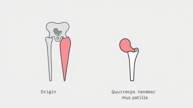
Origin of the Vastus Medialis Oblique
Primary Origin Sites
The VMO shares its origin with the larger vastus medialis muscle but has a specific fiber arrangement near the lower medial portion of the thigh. The general origin points of the VMO include:
- Medial lip of the linea aspera of the femur
- Medial supracondylar line of the femur
- Intermuscular septum
Fiber Orientation
What sets the VMO apart is the orientation of its muscle fibers. The oblique fibers of the VMO run at approximately a 5055 degree angle to the long axis of the femur, in contrast to the more vertical fibers of the upper vastus medialis. This angled alignment allows the VMO to exert a medial pull on the patella, counterbalancing the lateral pull from the vastus lateralis.
Insertion of the Vastus Medialis Oblique
Insertion Sites
The insertion of the VMO is into the quadriceps tendon and subsequently into the patella and tibial tuberosity via the patellar ligament. Specifically, the insertion includes:
- Medial border of the patella
- Quadriceps tendon, joining with fibers from the other quadriceps muscles
- Indirectly to the tibial tuberosity through the patellar ligament
This insertion structure allows the VMO to directly influence patellar tracking during knee extension and flexion.
Role in Patellar Tracking
The medial pull provided by the VMO at its insertion point helps guide the patella along the trochlear groove of the femur. Weakness or dysfunction in the VMO can result in lateral tracking of the patella, contributing to patellofemoral pain syndrome (PFPS), also known as runner’s knee.
Functional Importance of the VMO
Stabilization of the Patella
One of the VMO’s most crucial roles is to stabilize the patella medially, especially during the last 2030 degrees of knee extension. This function becomes particularly important in weight-bearing activities like walking, running, climbing stairs, and squatting.
Prevention of Knee Injuries
A strong and well-functioning VMO reduces the risk of common knee issues such as:
- Patellar subluxation or dislocation
- Patellofemoral pain syndrome
- Chondromalacia patellae
Rehabilitation programs often focus on targeting and strengthening the VMO in individuals with these conditions.
VMO Activation and Exercise
Challenges in Isolating the VMO
There is ongoing debate about whether the VMO can be activated independently of the other quadriceps muscles. While complete isolation may not be possible, certain exercises are believed to preferentially engage the VMO. These include:
- Terminal knee extensions (TKEs)
- Step-downs and step-ups
- Wall sits with a ball squeeze between the knees
- Mini-squats with hip adduction
Neuromuscular Control
Improving neuromuscular control of the VMO involves not just strength but timing and coordination. Delayed activation of the VMO relative to the vastus lateralis has been linked to patellofemoral dysfunction. Therefore, rehabilitation often includes both strengthening and retraining proper activation patterns.
Clinical Significance of VMO Anatomy
Assessment in Physical Therapy
Physical therapists assess the VMO’s function in patients with anterior knee pain, post-operative knee conditions, or athletic injuries. The origin and insertion points are critical landmarks for evaluating muscle tone, symmetry, and coordination.
Surgical and Post-Surgical Considerations
In surgical procedures such as total knee replacement or realignment surgeries, preserving or restoring VMO function is essential for optimal recovery. Surgeons may adjust techniques to protect the insertional region of the VMO and ensure proper tracking of the patella post-operatively.
Anatomical Variations and Controversies
Debate Over Distinct Muscle Status
Some anatomists argue whether the VMO is a separate muscle or merely a specialized part of the vastus medialis. Despite this debate, the clinical relevance of the VMO in patellar control is widely accepted, and its distinct oblique fiber orientation is universally recognized in functional anatomy.
Variability Among Individuals
The size, angle, and strength of the VMO vary between individuals. Athletes, especially those involved in sports requiring high degrees of knee control (like skiing, soccer, or basketball), often show well-developed VMO muscles. In contrast, sedentary individuals or those recovering from injury may display weakness or underdevelopment in this area.
The vastus medialis oblique is a critical component of the quadriceps group, distinguished by its unique origin on the medial aspect of the femur and its oblique fiber insertion into the patella via the quadriceps tendon. Its primary function is to medially stabilize the patella and assist in full knee extension. Dysfunction of the VMO is linked to several knee pathologies, making it an important focus in both injury prevention and rehabilitation. Understanding its anatomy particularly its origin and insertion is key for clinicians, therapists, and anyone involved in the care and training of the musculoskeletal system.