Congenital diaphragmatic hernia (CDH) is a rare but serious condition in which a defect in the diaphragm allows abdominal organs to move into the chest cavity, potentially interfering with lung development. This condition is usually identified in newborns, often presenting with respiratory distress shortly after birth. Timely diagnosis and intervention are critical to improving outcomes. While prenatal imaging like ultrasound and fetal MRI can sometimes detect CDH before birth, postnatal chest X-rays remain an essential diagnostic tool. Understanding the typical X-ray findings in congenital diaphragmatic hernia helps clinicians confirm diagnosis, plan surgical intervention, and monitor the newborn’s respiratory and cardiac status.
Role of X-Ray in Diagnosing Congenital Diaphragmatic Hernia
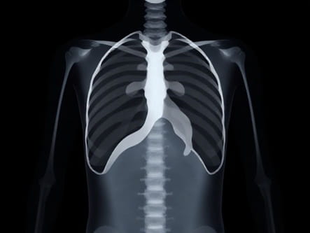
Chest X-rays are among the first imaging studies performed when CDH is suspected in a newborn. The X-ray provides a rapid, non-invasive method to visualize the thoracic cavity and detect abnormal positioning of abdominal organs. Radiographic evaluation helps confirm the diagnosis, determine the side of the hernia, and assess the degree of lung compression. In addition, X-rays can reveal associated complications such as mediastinal shift, pulmonary hypoplasia, or pleural effusion, which are critical for clinical management.
Typical X-Ray Findings in CDH
The characteristic features of congenital diaphragmatic hernia on chest X-ray depend on the location and size of the diaphragmatic defect, as well as which organs have herniated into the chest cavity.
- Bowel Loops in the ThoraxThe most common finding is gas-filled intestinal loops visible in the chest. These loops often appear as multiple cystic or tubular lucencies, commonly in the left hemithorax because left-sided CDH is more frequent.
- Mediastinal ShiftThe herniated organs push the mediastinum toward the opposite side of the chest. This shift may compromise cardiac function and exacerbate respiratory distress.
- Compressed LungsThe affected lung is usually hypoplastic and appears smaller with reduced aeration. This can be seen as increased density in the compressed lung field on X-ray.
- Absent or Elevated DiaphragmThe normal diaphragmatic contour may be absent or poorly defined. On some X-rays, the diaphragm appears higher on the affected side.
- Air-Fluid LevelsIf the stomach or intestines are herniated, air-fluid levels may be present, giving the appearance of multiple bubbles or lucent areas with fluid levels.
Types of Congenital Diaphragmatic Hernia and Their X-Ray Features
CDH is classified based on the location of the diaphragmatic defect. The most common types are Bochdalek and Morgagni hernias, each with distinct radiographic patterns.
Bochdalek Hernia
This type accounts for approximately 85% of CDH cases and typically occurs on the left posterior-lateral diaphragm. X-ray findings usually include
- Gas-filled bowel loops in the left thoracic cavity
- Mediastinal shift to the right
- Compression of the left lung with increased density
- Potential air-fluid levels if stomach or intestines are involved
Morgagni Hernia
Morgagni hernias are less common and occur in the anterior diaphragm near the sternum. X-ray features may include
- Opacity in the anterior mediastinum or lower lung fields
- Possible presence of bowel gas shadows if intestines herniate
- Less pronounced mediastinal shift compared to Bochdalek hernias
Complications Detected on X-Ray
Chest X-rays in CDH patients can also reveal complications that affect management decisions. Early recognition of these complications is critical for stabilizing newborns and planning surgery.
Pulmonary Hypoplasia
Compression of the lungs by herniated organs can lead to underdeveloped lung tissue. On X-ray, the hypoplastic lung appears smaller with increased opacity due to reduced aeration. Severe hypoplasia is associated with higher morbidity and mortality.
Mediastinal Shift
The displacement of the mediastinum, including the heart and great vessels, can be prominent on X-ray. Significant mediastinal shift may compromise cardiovascular stability and requires careful monitoring.
Pleural Effusions
Occasionally, pleural effusions may develop secondary to lung compression or cardiac compromise. These appear as fluid density along the chest wall or costophrenic angles on X-ray.
Limitations of X-Ray in CDH Diagnosis
Although X-rays are valuable for rapid assessment, they have limitations. They may not always clearly define the diaphragmatic defect, particularly in cases of small hernias or anterior Morgagni hernias. Overlapping structures can obscure bowel loops, making interpretation challenging. Furthermore, X-rays cannot provide detailed information about pulmonary vasculature or the degree of lung hypoplasia, which may necessitate additional imaging such as ultrasound, CT, or MRI for comprehensive evaluation.
Role of X-Ray in Preoperative and Postoperative Assessment
X-ray imaging is essential not only for initial diagnosis but also for monitoring before and after surgical repair of CDH. Preoperative X-rays help assess lung compression, mediastinal shift, and presence of herniated organs, guiding stabilization and surgical planning. Postoperative X-rays are used to evaluate the position of repaired organs, the integrity of the diaphragm repair, and detect potential complications like pneumothorax, residual pleural effusions, or recurrent herniation.
Integration with Other Diagnostic Tools
While X-ray provides initial and ongoing assessment, comprehensive management of CDH often requires integration with other imaging modalities
- UltrasoundHelps assess abdominal organ position, lung volume, and presence of associated anomalies.
- CT ScanOffers detailed anatomical information and can evaluate diaphragmatic defect size and surrounding structures.
- MRIUseful in prenatal assessment, providing information on lung-to-head ratio and predicting postnatal outcomes.
Chest X-rays play a pivotal role in diagnosing congenital diaphragmatic hernia, offering rapid visualization of herniated organs, lung compression, mediastinal shift, and other complications. Recognizing typical X-ray patterns is essential for timely intervention, stabilization of the newborn, and surgical planning. Despite its limitations, X-ray remains an indispensable tool in the evaluation of CDH, especially when integrated with other imaging modalities. Proper interpretation of X-ray findings ensures accurate diagnosis, guides clinical decision-making, and helps improve outcomes in affected infants.