The greater occipital nerve is a significant structure in human anatomy, responsible for providing sensation to a large portion of the posterior scalp. Studying this nerve through cadaver dissection is essential for medical students, clinicians, and researchers aiming to understand its anatomical course, variations, and clinical implications. The greater occipital nerve emerges from the dorsal ramus of the second cervical spinal nerve (C2) and travels through muscles and connective tissue to innervate the scalp. Examining the nerve in cadaver specimens allows for precise observation of its trajectory, relationships with surrounding structures, and potential sites for clinical intervention, such as nerve blocks used to treat occipital neuralgia.
Anatomical Overview of the Greater Occipital Nerve
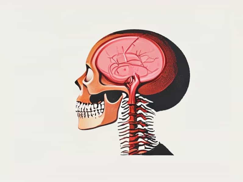
The greater occipital nerve originates from the dorsal ramus of C2, sometimes receiving contributions from C3. After exiting between the atlas and axis vertebrae, the nerve travels superiorly, passing beneath or through the semispinalis capitis and trapezius muscles before reaching the scalp. Along its course, it provides sensory innervation to the posterior scalp, extending from the base of the skull to the vertex. The nerve is often accompanied by branches of the occipital artery, which can serve as landmarks during anatomical dissection and clinical procedures.
Cadaver Dissection Techniques
Dissecting the greater occipital nerve in cadaver specimens requires careful attention to detail to preserve the integrity of the nerve and its surrounding structures. Typically, the dissection begins with an incision along the posterior neck, exposing the suboccipital muscles. The trapezius and semispinalis capitis are carefully reflected to reveal the nerve. Identifying the nerve’s origin at C2 and tracing its course superiorly provides valuable insight into anatomical variations, branching patterns, and relationships with adjacent muscles and vascular structures. Cadaver studies often reveal slight differences between individuals, emphasizing the importance of understanding anatomical variability for surgical planning and pain management procedures.
Clinical Relevance of the Greater Occipital Nerve
The greater occipital nerve is clinically important due to its involvement in occipital neuralgia, a condition characterized by chronic pain in the back of the head and neck. Compression or irritation of the nerve can result from trauma, muscle tightness, or degenerative cervical spine conditions. Cadaver studies provide critical anatomical knowledge that informs nerve block techniques, where local anesthetic is injected near the nerve to relieve pain. Understanding the exact course of the nerve, its relationship to the occipital artery, and variations in branching is essential for performing these procedures safely and effectively.
Surgical and Diagnostic Applications
In addition to pain management, knowledge of the greater occipital nerve anatomy is crucial in surgical procedures involving the posterior cervical region. Surgeons must avoid nerve injury during procedures such as suboccipital craniectomies, posterior cervical fusions, and occipital artery bypasses. Cadaveric studies allow surgeons to practice dissections, identify key landmarks, and anticipate potential variations. Diagnostic procedures, including nerve stimulation and imaging studies, also benefit from an in-depth understanding of the nerve’s anatomical course as observed in cadaver specimens.
Variations and Anatomical Considerations
Anatomical studies of cadavers often reveal variations in the greater occipital nerve. These variations may include differences in the number of branches, the nerve’s relationship to surrounding muscles, and its proximity to the occipital artery. Some individuals may have a more medial or lateral nerve trajectory, which can influence the effectiveness of nerve blocks and surgical approaches. Cadaveric research provides a comprehensive view of these variations, allowing clinicians to tailor interventions based on individual anatomical differences, thereby increasing safety and efficacy.
Educational Importance
Studying the greater occipital nerve in cadaver specimens is an invaluable educational tool for medical students and residents. Hands-on dissection helps students visualize three-dimensional relationships, understand anatomical variability, and develop a tactile appreciation for the tissue planes surrounding the nerve. This experience enhances their ability to apply anatomical knowledge in clinical settings, whether performing nerve blocks, diagnosing occipital neuralgia, or planning surgical procedures. Cadaveric studies bridge the gap between textbook diagrams and real-life anatomy, fostering a deeper understanding of neuroanatomy.
Techniques for Preserving Nerve Integrity in Cadaver Studies
- Use fine dissection tools to carefully separate the greater occipital nerve from surrounding muscles and connective tissue.
- Identify the occipital artery early in dissection as a landmark to trace the nerve’s path.
- Document variations in branching patterns and muscle relationships for reference in clinical practice.
- Maintain proper preservation techniques to avoid distortion of tissue planes and nerve structures.
- Practice incremental dissection, tracing the nerve from its origin at C2 to its terminal branches on the scalp.
The greater occipital nerve is a critical anatomical structure with significant clinical and educational relevance. Studying the nerve in cadaver specimens allows for detailed observation of its course, branching patterns, and relationships with surrounding muscles and vessels. Such knowledge is essential for diagnosing and managing occipital neuralgia, performing safe surgical procedures, and enhancing the anatomical education of medical professionals. Cadaveric dissections provide a practical and visual approach to understanding the greater occipital nerve, bridging theoretical knowledge and clinical application. Through these studies, clinicians and students gain insight into anatomical variability, refine surgical techniques, and improve patient outcomes in conditions involving the posterior scalp and cervical region.