The greater trochanter of the femur is one of the most important bony landmarks of the human body, located on the upper part of the thigh bone. It plays a vital role in connecting muscles, facilitating hip movement, and maintaining balance during standing, walking, or running. This structure is easily palpable through the skin on the outer side of the hip, and it serves as an essential reference point for both medical professionals and students of anatomy. Understanding the greater trochanter helps explain how the hip joint functions as one of the strongest and most stable joints in the body.
Anatomical Location of the Greater Trochanter
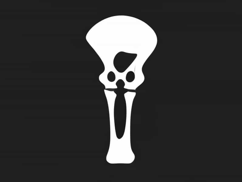
The greater trochanter is a large, irregularly shaped projection situated on the lateral side of the femur, near the top of the thigh bone. It extends from the junction of the neck and shaft of the femur. If you touch the side of your hip, you can feel a hard prominence under the skin that is the greater trochanter. It lies opposite to another smaller projection called the lesser trochanter, which is located on the inner side and slightly lower.
This structure is part of the proximal femur, which includes the femoral head, neck, greater trochanter, lesser trochanter, and the intertrochanteric line and crest. Together, these structures form the upper part of the femur that articulates with the pelvis at the hip joint. The greater trochanter provides attachment points for several important muscles that control movement of the hip and thigh.
Muscular Attachments
One of the main functions of the greater trochanter is to serve as an anchor for muscles that stabilize and move the hip joint. Several muscles attach to different surfaces of the greater trochanter, contributing to movements such as abduction, rotation, and stabilization of the thigh.
Muscles Attached to the Greater Trochanter
- Gluteus mediusAttaches to the lateral surface of the greater trochanter and is responsible for abducting the thigh, moving it away from the body’s midline.
- Gluteus minimusAttaches to the anterior surface of the greater trochanter and assists in both abduction and medial rotation of the hip.
- PiriformisInserts on the upper border of the greater trochanter and helps in lateral rotation of the thigh.
- Obturator internusInserts on the medial surface, also aiding in lateral rotation of the hip.
- Gemellus superior and inferiorThese small muscles insert near the obturator internus tendon, contributing to hip stabilization and rotation.
Together, these muscles help maintain the position of the pelvis during walking and standing on one leg. The balance between these muscle groups is essential for smooth and coordinated lower limb movements.
Structure and Surfaces
The greater trochanter has multiple surfaces and borders that serve specific anatomical purposes. Each surface is associated with muscle attachments and functions.
- Lateral surfaceBroad and convex, providing insertion for the gluteus medius muscle.
- Medial surfaceContains a deep pit known as the trochanteric fossa, where the obturator internus and gemelli muscles attach.
- Anterior surfaceSmooth area that serves as the site of attachment for the gluteus minimus muscle.
- Superior borderThe top edge where the piriformis muscle attaches.
- Posterior borderRough and irregular, forming part of the insertion area for the gluteal muscles.
The bone itself is dense and strong, designed to withstand the tension and pressure generated by the powerful muscles surrounding the hip joint. The arrangement of the greater trochanter allows for efficient transfer of force from the muscles to the femur during movement.
Function and Biomechanics
The greater trochanter plays a crucial role in lower limb biomechanics. It acts as a lever for the muscles attached to it, enhancing their mechanical advantage. During activities such as walking, climbing stairs, or running, the greater trochanter functions as a point of rotation that allows for smooth hip movement.
When a person walks, the gluteus medius and minimus muscles contract to keep the pelvis level on the opposite side. Without the support of the greater trochanter, these muscles would have reduced leverage, making it difficult to maintain balance. Additionally, the greater trochanter helps distribute body weight evenly across the hip joint, preventing strain and promoting efficient motion.
Clinical Significance
The greater trochanter of the femur is often involved in medical conditions related to the hip. Because of its exposed position and muscular attachments, it can be affected by trauma, overuse, or inflammation. Understanding these conditions is important for diagnosis and treatment.
Common Conditions Affecting the Greater Trochanter
- Trochanteric bursitisInflammation of the bursa overlying the greater trochanter. It causes pain on the outer side of the hip, often worsened by walking or lying on the affected side.
- Greater trochanteric pain syndrome (GTPS)A broader term that includes bursitis, tendon inflammation, and muscle tears around the greater trochanter.
- FracturesThe greater trochanter can be fractured in falls or direct impacts, especially in elderly individuals with osteoporosis. Such fractures may occur alone or as part of a femoral neck fracture.
- TendinopathyOveruse of the hip muscles, particularly in athletes or runners, can cause inflammation or degeneration of tendons attached to the greater trochanter.
These conditions typically cause lateral hip pain, tenderness to touch, and difficulty moving the hip. Treatments may include rest, physical therapy, anti-inflammatory medications, and in severe cases, surgical intervention.
Diagnostic and Imaging Importance
The greater trochanter serves as a useful landmark in diagnostic imaging and surgical procedures. On X-rays or MRI scans, it is often used to assess hip alignment, muscle attachment integrity, and fracture locations. In orthopedic surgery, it acts as a key reference point for hip replacement operations, ensuring accurate placement of prosthetic components.
Additionally, palpation of the greater trochanter helps clinicians evaluate hip alignment, muscle tenderness, and range of motion. Because it is easily accessible, it is frequently used during physical examinations to locate pain or swelling around the hip region.
Development and Growth
During growth, the greater trochanter develops from a separate ossification center. In children, it is primarily made of cartilage that gradually ossifies as they mature. Complete fusion with the femoral shaft typically occurs in late adolescence. Proper alignment during growth is important, as abnormal development can lead to altered hip mechanics and long-term functional problems.
Relation to Surrounding Structures
Surrounding the greater trochanter are several soft tissues, including tendons, bursae, and fascia. The trochanteric bursa, located between the greater trochanter and overlying muscles, helps reduce friction during movement. The iliotibial band also runs close to the greater trochanter, providing lateral stability to the thigh. These structures work together to support and protect the hip joint during activity.
The greater trochanter of the femur is far more than just a bony prominence on the side of the thigh. It is a vital structural and functional component of the hip joint, serving as the attachment site for key muscles that control leg movement and maintain balance. Its importance extends from anatomy and biomechanics to clinical medicine, where it plays a role in diagnosing and treating hip pain and injury. Understanding this part of the femur gives deeper insight into how the human body achieves stability, strength, and smooth movement in everyday life.