The human urinary system is a complex network responsible for filtering blood, producing urine, and maintaining the body’s fluid and electrolyte balance. Understanding the structure and function of the kidneys, ureters, and bladder is crucial for both medical education and general health awareness. A kidney ureter bladder diagram, often abbreviated as a KUB diagram, visually represents these organs, their relationships, and their roles in excreting waste. Such diagrams are widely used in anatomy classes, medical imaging studies, and patient education, providing a clear overview of how urine travels from the kidneys to the bladder for storage before elimination. Learning about this system helps people understand common urinary disorders and promotes better health practices.
Overview of the Kidney, Ureter, and Bladder
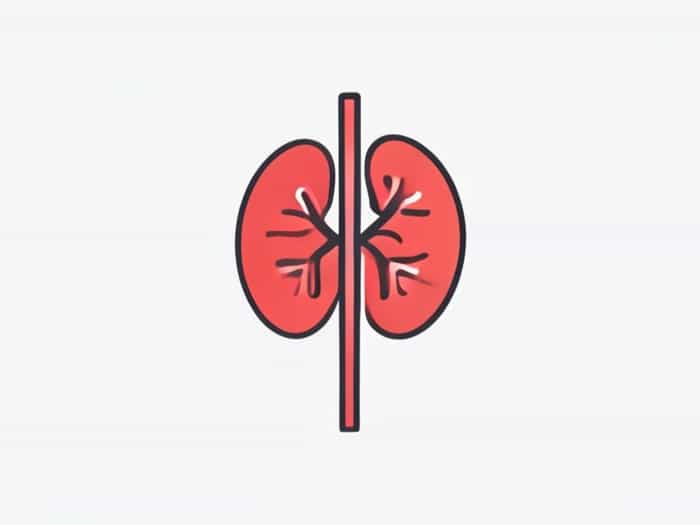
The urinary system is composed primarily of the kidneys, ureters, and bladder, each with a distinct function. The kidneys are bean-shaped organs located on either side of the spine, responsible for filtering blood, removing waste, and balancing electrolytes. The ureters are narrow tubes that transport urine from the kidneys to the bladder. The bladder is a muscular sac that stores urine until it is expelled from the body. Together, these organs form a continuous pathway for urine flow and are crucial for maintaining homeostasis. A kidney ureter bladder diagram simplifies this anatomy, helping learners visualize the connections and functions of these organs.
Kidneys Structure and Function
The kidneys are vital organs that play a central role in filtering waste from the bloodstream. Each kidney contains around one million nephrons, which are microscopic units that filter blood, reabsorb necessary substances, and secrete waste as urine. In addition to excretion, kidneys regulate blood pressure, produce hormones like erythropoietin, and maintain fluid and electrolyte balance. The kidney ureter bladder diagram typically shows the kidneys located on either side of the vertebral column, highlighting their shape, position, and connection to the ureters.
Ureters Pathways for Urine
The ureters are muscular tubes that carry urine from the kidneys to the bladder. Each ureter is about 25-30 centimeters long and uses peristaltic contractions to move urine efficiently. The ureters enter the bladder at an angle, which helps prevent urine from flowing backward, reducing the risk of infection. In a kidney ureter bladder diagram, the ureters are usually depicted as slender tubes connecting the renal pelvis of each kidney to the bladder, showing their critical role in urine transport.
Bladder Storage and Release of Urine
The bladder is a hollow, muscular organ located in the pelvic cavity that stores urine until it is convenient to release it. The bladder walls are composed of a specialized muscle called the detrusor, which expands as the bladder fills and contracts during urination. The bladder has two main openings for the ureters and one opening for the urethra, through which urine exits the body. In a kidney ureter bladder diagram, the bladder is typically shown as a rounded structure at the base of the urinary tract, illustrating its function as a temporary reservoir.
Kidney Ureter Bladder Diagram in Medical Education
Medical students, healthcare professionals, and educators often use a kidney ureter bladder diagram to simplify the study of urinary anatomy. The diagram helps learners visualize organ placement, understand the flow of urine, and identify areas prone to medical conditions such as kidney stones, urinary tract infections, or blockages. Detailed KUB diagrams may include labels for renal arteries, veins, the renal pelvis, and the internal structure of the bladder, offering a comprehensive view of urinary anatomy. By using a diagram, students can quickly grasp the relationships between the kidneys, ureters, and bladder, which is essential for understanding physiology and pathology.
Clinical Relevance of a KUB Diagram
The kidney ureter bladder diagram is not only an educational tool but also has significant clinical applications. In radiology, KUB X-rays are commonly performed to detect stones, obstructions, or other abnormalities in the urinary tract. By comparing the X-ray images to the standard KUB diagram, clinicians can identify deviations from normal anatomy and diagnose conditions accurately. Additionally, understanding the normal anatomy through diagrams helps surgeons plan procedures, such as ureteral stent placement or bladder surgery. Patients can also benefit from visual explanations using KUB diagrams, which enhance understanding of their condition and treatment options.
Common Urinary Conditions Illustrated by KUB Diagrams
KUB diagrams help in visualizing several common urinary system conditions
- Kidney StonesHard mineral deposits that form in the kidneys and may travel through the ureters to the bladder.
- Urinary Tract InfectionsInfections that can affect the bladder, ureters, or kidneys, often visualized by swelling or obstruction in diagrams.
- HydronephrosisSwelling of a kidney due to urine buildup, often identified in imaging studies alongside diagrams.
- Congenital AbnormalitiesStructural differences in the urinary tract, which can be better understood with diagrams.
Advantages of Using Kidney Ureter Bladder Diagrams
Visual diagrams provide several advantages for learning and clinical practice. They simplify complex anatomical relationships, making it easier to understand the urinary system’s function. Diagrams also assist in memorization, enabling students and practitioners to recall organ positions and connections quickly. Furthermore, diagrams are valuable in patient education, allowing doctors to explain conditions, procedures, and treatment plans more effectively. For radiologists and surgeons, KUB diagrams serve as reference points to compare normal anatomy with imaging results, improving diagnostic accuracy.
Tips for Interpreting KUB Diagrams
- Identify the kidneys first, noting their shape, position, and orientation.
- Trace the ureters from the renal pelvis to the bladder, observing their curvature and path.
- Examine the bladder at the base of the diagram, noting its size and muscular structure.
- Compare the diagram with imaging studies to detect anomalies or blockages.
A kidney ureter bladder diagram is an essential tool for understanding the human urinary system. By visually representing the kidneys, ureters, and bladder, it provides clarity on organ function, relationships, and urine flow. The diagram is invaluable for students, healthcare professionals, and patients, helping to explain anatomy, diagnose conditions, and plan treatments. Additionally, it plays a key role in identifying common urinary problems such as stones, infections, and structural abnormalities. Learning through diagrams not only enhances comprehension of urinary anatomy but also fosters better health awareness and medical decision-making. Whether in the classroom, clinic, or home setting, the KUB diagram serves as a foundational reference for understanding one of the body’s most vital systems.