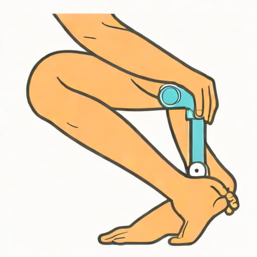
Knee extension is a fundamental movement that plays a crucial role in daily activities such as walking, running, standing up, and climbing stairs. Understanding the innervation of the muscles responsible for knee extension is essential for students, healthcare professionals, and anyone interested in anatomy or rehabilitation. The process involves a complex interaction between the nervous system and the muscles of the anterior thigh, primarily the quadriceps femoris group. Proper innervation ensures coordinated movement, strength, and stability of the knee joint. Any disruption in nerve supply can lead to weakness, impaired function, or even complete loss of knee extension, significantly affecting mobility and quality of life. Studying knee extension innervation provides insights into clinical conditions such as nerve injuries, neuropathies, and post-surgical rehabilitation strategies.
Anatomy of Knee Extension
Knee extension is mainly performed by the quadriceps femoris, a large muscle group located in the anterior compartment of the thigh. The quadriceps consist of four distinct muscles rectus femoris, vastus lateralis, vastus medialis, and vastus intermedius. Each muscle has a specific origin and insertion, but all converge into the quadriceps tendon, which attaches to the patella and continues as the patellar ligament to the tibial tuberosity. The rectus femoris also crosses the hip joint, contributing to hip flexion. The combined action of these muscles straightens the knee from a flexed position and provides stability during weight-bearing activities.
Muscles Involved in Knee Extension
- Rectus Femoris Originates from the anterior inferior iliac spine and contributes to both knee extension and hip flexion.
- Vastus Lateralis Originates from the greater trochanter and lateral lip of the linea aspera, primarily responsible for lateral knee stability during extension.
- Vastus Medialis Originates from the intertrochanteric line and medial lip of the linea aspera, crucial for medial knee stability and patellar tracking.
- Vastus Intermedius Located deep to the rectus femoris, originates from the anterior and lateral surfaces of the femur, assisting in knee extension.
Nervous System and Innervation
The innervation of knee extension muscles is primarily provided by the femoral nerve, a major nerve of the lumbar plexus. The femoral nerve arises from the posterior divisions of the ventral rami of the L2, L3, and L4 spinal nerves. It travels through the pelvis, passing beneath the inguinal ligament to enter the anterior thigh compartment. Once in the thigh, the femoral nerve branches into several motor and sensory nerves, with motor branches specifically targeting the quadriceps femoris muscles. These motor fibers transmit electrical signals from the spinal cord, triggering muscle contraction and enabling knee extension.
Femoral Nerve Pathway
The femoral nerve is the largest branch of the lumbar plexus and plays a critical role in both motor and sensory functions of the anterior thigh. After exiting the lumbar plexus, it descends alongside the psoas major muscle and passes under the inguinal ligament to reach the femoral triangle. Here, the nerve divides into multiple branches
- Muscular branches Supply the quadriceps femoris and sartorius muscles, enabling knee extension and hip flexion.
- Saphenous nerve Provides sensory innervation to the medial aspect of the leg and foot.
Through these branches, the femoral nerve ensures that the quadriceps receive the necessary stimulation for efficient knee extension.
Clinical Significance of Knee Extension Innervation
Proper innervation of the knee extensor muscles is essential for maintaining mobility, stability, and functional independence. Damage or impairment of the femoral nerve can result in weakness or paralysis of the quadriceps, leading to difficulty in standing up from a seated position, climbing stairs, or walking efficiently. Common causes of femoral nerve dysfunction include traumatic injuries, compression due to tumors, iatrogenic injury during surgery, and neuropathies. Assessing the strength and function of knee extension is a key part of neurological examinations and rehabilitation planning.
Assessment of Quadriceps Function
Clinicians evaluate knee extension strength through physical examination techniques such as manual muscle testing, where resistance is applied to assess quadriceps force. Reflex testing, including the patellar tendon reflex, helps determine the integrity of the femoral nerve and the L2-L4 spinal segments. Abnormal results may indicate nerve injury, spinal cord pathology, or muscular disorders, guiding further diagnostic and therapeutic interventions.
Rehabilitation and Therapy
Rehabilitation following femoral nerve injury or quadriceps weakness focuses on restoring muscle strength, joint stability, and functional mobility. Treatment strategies may include
- Physical therapy exercises to strengthen the quadriceps, such as straight leg raises and seated knee extensions.
- Electrical stimulation to facilitate muscle contraction in cases of partial nerve damage.
- Gait training and balance exercises to improve walking and reduce fall risk.
- Orthotic devices, such as knee braces, to support weakened muscles during recovery.
Early intervention and consistent therapy improve outcomes and help patients regain independence in daily activities.
Integration with Other Muscle Groups
Knee extension does not occur in isolation; it is integrated with other muscle groups to achieve coordinated lower limb movement. The hamstrings, located at the posterior thigh, act as antagonists during knee extension, controlling the speed and smoothness of the movement. Additionally, hip flexors, gluteal muscles, and calf muscles work synergistically during activities like running and jumping. Understanding the innervation of knee extension in the context of surrounding muscles is essential for comprehensive rehabilitation and performance optimization.
Neurological Control and Reflexes
The nervous system regulates knee extension through both voluntary and reflexive pathways. Voluntary movement originates in the motor cortex, sending signals via corticospinal tracts to the femoral nerve. Reflexive actions, such as the knee-jerk reflex, are mediated by sensory and motor fibers connecting the muscle spindle to the spinal cord, providing rapid adjustments to maintain posture and stability. These mechanisms ensure precise, controlled knee extension during dynamic activities.
Knee extension innervation is a complex and vital aspect of human movement, primarily involving the quadriceps femoris and the femoral nerve. Proper function of these muscles and their nerve supply is essential for mobility, stability, and overall quality of life. Understanding the anatomy, innervation pathways, clinical significance, and rehabilitation strategies provides valuable insights for healthcare professionals, athletes, and students of anatomy. Disruption in nerve function can lead to significant impairment, highlighting the importance of early diagnosis, targeted therapy, and careful monitoring. By integrating knowledge of knee extension innervation with broader musculoskeletal and neurological principles, we can optimize patient care, enhance performance, and support recovery in cases of injury or disease.