The congruity of the hip joint is a fundamental aspect of orthopedic health, biomechanics, and overall mobility. It refers to the precise alignment and complementary surfaces of the femoral head and the acetabulum, which allow smooth motion, effective load distribution, and long-term joint stability. Proper congruity ensures that forces generated during activities such as walking, running, or jumping are evenly transmitted across the joint surfaces, minimizing wear and tear and preventing degenerative changes. Understanding hip joint congruity is essential for clinicians, physical therapists, and patients alike, as it underpins the diagnosis, treatment, and prevention of various hip pathologies, including osteoarthritis, dysplasia, and labral injuries.
Anatomy of the Hip Joint
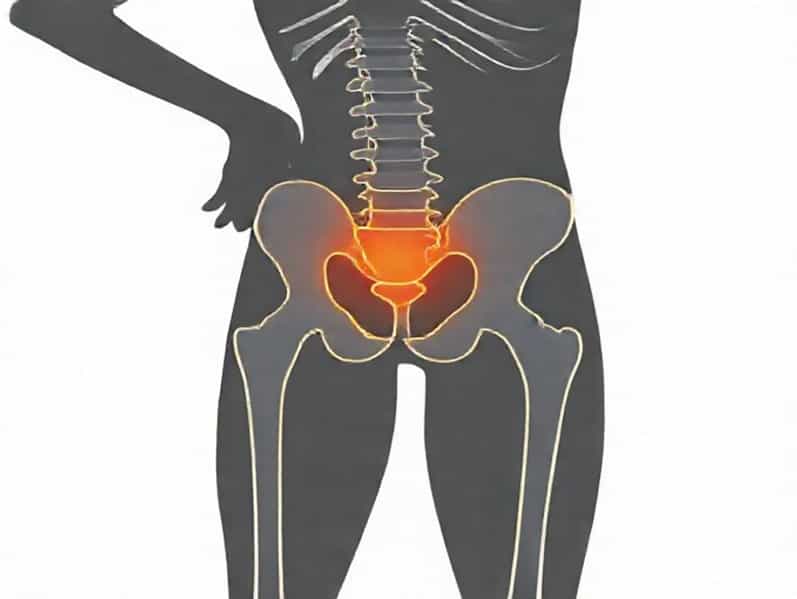
The hip joint is a ball-and-socket synovial joint, where the spherical femoral head fits into the concave acetabulum of the pelvis. This unique structure provides a wide range of motion while maintaining stability. The articular cartilage lining both the femoral head and acetabulum contributes to congruity by creating a smooth, frictionless surface for movement. The labrum, a fibrocartilaginous rim surrounding the acetabulum, enhances joint stability and deepens the socket, further supporting congruity. Surrounding muscles, ligaments, and tendons maintain joint alignment and facilitate functional movement.
Factors Affecting Hip Joint Congruity
Several anatomical and physiological factors influence the congruity of the hip joint, including
- Acetabular DepthA deeper acetabulum generally enhances congruity by securely accommodating the femoral head, whereas a shallow socket may predispose to instability.
- Femoral Head ShapeA perfectly spherical femoral head contributes to optimal congruity. Deformities such as asphericity can reduce contact area and increase stress on the cartilage.
- Cartilage IntegrityHealthy cartilage ensures even load distribution and smooth articulation. Degeneration or thinning can disrupt congruity and lead to pain and restricted movement.
- Labral FunctionThe labrum helps maintain a suction effect that stabilizes the femoral head. Labral tears or degeneration can compromise congruity.
Biomechanics and Function
Hip joint congruity is essential for efficient biomechanics. A congruent joint allows even distribution of mechanical forces across the articular surfaces, reducing peak stress points that can lead to microdamage. Proper congruity also facilitates smooth rotation, flexion, extension, abduction, and adduction without causing joint impingement or instability. When congruity is compromised, abnormal stress patterns develop, predisposing the joint to conditions such as osteoarthritis, labral tears, and femoroacetabular impingement.
Load Transmission
During weight-bearing activities, the hip joint transmits forces from the upper body to the lower limbs. In a congruent joint, these forces are evenly distributed, preventing excessive wear on the cartilage. Studies have shown that minor incongruities, even if asymptomatic, can increase peak contact stress, accelerating cartilage degeneration over time. Therefore, maintaining congruity is crucial for joint longevity, especially in physically active individuals or athletes.
Range of Motion
The spherical nature of the femoral head and the concave acetabulum allows multi-directional movement. Congruity ensures that motion is smooth and unrestricted. Loss of congruity, due to deformities, dysplasia, or injury, may lead to abnormal kinematics, restricted motion, and compensatory gait patterns. Early recognition and intervention can help restore function and prevent secondary musculoskeletal problems.
Assessment of Hip Joint Congruity
Evaluating hip congruity involves clinical examination and imaging studies. Clinicians look for symmetry, joint space, and alignment, while patients may report pain, instability, or restricted motion. Imaging techniques, such as X-rays, MRI, or CT scans, provide detailed views of the femoral head, acetabulum, cartilage, and labrum. Radiographic measures, including the center-edge angle, acetabular index, and joint space width, help quantify congruity and identify abnormalities that may require treatment.
Clinical Indicators
- Pain during weight-bearing activities or at rest
- Reduced range of motion, particularly in rotation or flexion
- Instability or a feeling of the hip giving way
- Clicking, catching, or locking sensations indicating labral or cartilage issues
Imaging Techniques
X-rays are often the first step in assessing hip joint congruity, revealing joint space narrowing, subluxation, or acetabular dysplasia. MRI provides detailed visualization of cartilage, labrum, and soft tissues, which is essential for diagnosing early degenerative changes or labral tears. CT scans offer three-dimensional assessment of bony structures, helpful in preoperative planning for corrective procedures or joint replacement.
Pathological Conditions Affecting Congruity
Several conditions can disrupt the congruity of the hip joint, leading to pain, reduced function, and degenerative changes. Early detection and intervention are crucial to prevent long-term damage. Common conditions include
Hip Dysplasia
Developmental dysplasia of the hip involves a shallow acetabulum or malaligned femoral head, resulting in poor congruity. If untreated, it can lead to early osteoarthritis and gait abnormalities. Treatment may involve bracing in infants or surgical procedures in older children and adults.
Osteoarthritis
Degenerative joint disease reduces cartilage thickness, alters femoral head shape, and causes subchondral bone changes. These factors compromise congruity and increase stress on remaining cartilage, perpetuating a cycle of degeneration and pain.
Femoroacetabular Impingement
Abnormal bony growths on the femoral head or acetabulum can lead to impingement during movement, disrupting congruity. Patients often experience pain, restricted motion, and labral tears. Surgical intervention may restore congruity and preserve joint function.
Labral Tears
The labrum stabilizes the femoral head and maintains congruity. Tears due to trauma or repetitive stress reduce joint stability and can accelerate cartilage wear. Arthroscopic repair or debridement may be necessary to restore function.
Maintaining and Restoring Hip Congruity
Preserving or restoring hip joint congruity is key to long-term mobility and quality of life. Strategies include
- Early diagnosis and treatment of congenital or developmental disorders
- Physical therapy to strengthen surrounding muscles and maintain alignment
- Minimally invasive surgery to correct deformities or repair labral injuries
- Joint replacement in advanced osteoarthritis to restore congruent articulating surfaces
Rehabilitation and Exercise
Targeted exercises help maintain muscle balance, joint stability, and proper alignment. Strengthening the gluteal, quadriceps, and hip rotator muscles supports the femoral head within the acetabulum, reducing abnormal stress and preserving congruity. Stretching and mobility exercises improve flexibility, allowing smooth joint motion and reducing the risk of impingement or injury.
The congruity of the hip joint is a critical determinant of musculoskeletal health, functional mobility, and long-term joint integrity. Proper alignment and complementary surfaces between the femoral head and acetabulum ensure even load distribution, smooth motion, and stability. Disruptions in congruity, whether due to congenital, developmental, or degenerative conditions, can lead to pain, impaired movement, and early joint deterioration. Assessment through clinical examination and imaging is essential for early detection, and interventions such as physical therapy, corrective surgery, or joint replacement can restore or preserve congruity. Understanding hip joint congruity not only benefits clinicians in diagnosis and treatment but also helps individuals recognize the importance of maintaining hip health through exercise, injury prevention, and proactive care.