A fetal anatomic survey ultrasound is a crucial diagnostic tool used in prenatal care to evaluate the development and well-being of a fetus during pregnancy. This type of ultrasound, often performed between 18 and 22 weeks of gestation, allows healthcare providers to assess fetal anatomy in detail, identify structural abnormalities, and monitor growth and development. The procedure provides essential information that helps guide medical decisions, plan for delivery, and prepare for potential interventions if abnormalities are detected. Understanding the purpose, procedure, and significance of a fetal anatomic survey ultrasound is important for expectant parents and healthcare providers alike.
Purpose of Fetal Anatomic Survey Ultrasound
The main purpose of a fetal anatomic survey ultrasound is to evaluate the fetus for structural and developmental anomalies. By examining the organs, limbs, and overall body structure, clinicians can detect potential health concerns early. Early detection allows for timely interventions, specialized care, or additional testing if necessary.
Evaluation of Fetal Anatomy
During the ultrasound, detailed images are captured of key fetal structures. These include the brain, heart, spine, abdomen, kidneys, bladder, stomach, limbs, and facial features. Each organ is assessed for size, shape, position, and overall integrity. Any abnormalities, such as heart defects, neural tube defects, or limb malformations, can be identified during this comprehensive examination.
Monitoring Growth and Development
The fetal anatomic survey also measures growth parameters, including head circumference, abdominal circumference, and femur length. These measurements help determine if the fetus is developing appropriately for gestational age. Consistent growth monitoring is important for detecting conditions such as intrauterine growth restriction or macrosomia, which may impact the health of the baby and the management of the pregnancy.
Timing of the Ultrasound
The optimal timing for a fetal anatomic survey ultrasound is typically between 18 and 22 weeks of pregnancy. At this stage, the fetus is large enough to allow detailed imaging, and the organs have developed sufficiently for evaluation. Performing the survey within this window maximizes the likelihood of identifying anomalies while still allowing time for medical decision-making if interventions are needed.
First vs. Second Trimester Screening
While initial screening ultrasounds may occur in the first trimester to confirm pregnancy and estimate gestational age, the detailed fetal anatomic survey is conducted in the second trimester. First trimester ultrasounds can detect some major abnormalities, but the second trimester survey provides a more comprehensive assessment of fetal structures and organ development.
Procedure of Fetal Anatomic Survey Ultrasound
The fetal anatomic survey ultrasound is a non-invasive procedure that uses high-frequency sound waves to create images of the developing fetus. The procedure is generally safe and painless for both the mother and the fetus, and it does not involve radiation.
Preparation for the Ultrasound
Patients are usually advised to drink water before the procedure to ensure a full bladder, which improves visualization of the uterus and fetus. Comfortable clothing and prior medical records may also be helpful for the ultrasound technician or physician conducting the exam.
Conducting the Ultrasound
During the procedure, the patient lies on an examination table while a transducer is moved across the abdomen. The transducer emits sound waves that bounce off fetal tissues, creating images on a monitor. The sonographer or physician captures multiple images of the fetus from different angles to ensure that all organs and structures are thoroughly examined.
Use of Doppler Imaging
In some cases, Doppler imaging may be used to assess blood flow in fetal vessels, the placenta, and the umbilical cord. This evaluation can provide additional information about fetal health, including oxygenation and nutrient delivery, which are critical for growth and development.
Key Structures Evaluated
A fetal anatomic survey ultrasound examines multiple organ systems to ensure proper development. Each system is carefully evaluated for abnormalities or variations from normal growth patterns.
Brain and Central Nervous System
The brain and spinal cord are assessed for size, structure, and proper formation. This includes checking for conditions such as ventriculomegaly, spina bifida, or other neural tube defects. Cranial and facial structures are also examined for abnormalities that could affect development.
Cardiac Assessment
The fetal heart is evaluated for proper chamber formation, valve function, and blood flow. Congenital heart defects, which are among the most common birth anomalies, can often be detected during this examination. The sonographer may use Doppler imaging to assess circulation and identify potential issues early.
Abdominal and Genitourinary Organs
The stomach, kidneys, bladder, and other abdominal organs are checked for size, position, and proper function. The survey ensures that the digestive and urinary systems are developing normally and identifies any obstructions or malformations.
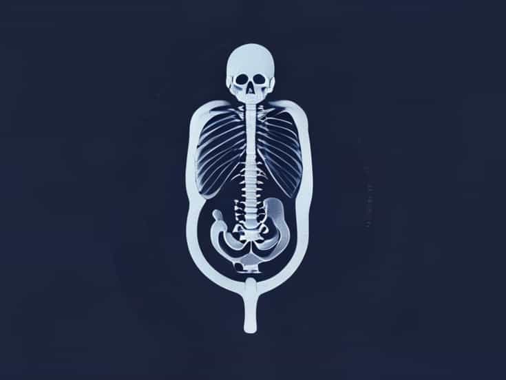
Limb and Skeletal Structures
All limbs, hands, feet, and joints are examined for proper formation. Measurements are taken to ensure that growth is proportional. The spine is also carefully evaluated to detect abnormalities such as scoliosis or other skeletal defects.
Placenta and Umbilical Cord
The position, structure, and function of the placenta are assessed, along with the umbilical cord. The sonographer looks for signs of placenta previa, placental insufficiency, or cord abnormalities, which may affect fetal growth or complicate delivery.
Importance of the Fetal Anatomic Survey Ultrasound
This detailed ultrasound provides critical information for managing pregnancy and preparing for potential challenges. Early identification of abnormalities allows for counseling, further testing, or interventions to improve outcomes for both the mother and the baby.
Guiding Medical Decisions
Information from the fetal anatomic survey can influence decisions about prenatal care, delivery planning, and the need for additional diagnostic procedures. For example, detecting a structural anomaly may prompt genetic testing, specialist consultations, or early interventions to address complications before birth.
Reassurance for Parents
For expectant parents, a normal fetal anatomic survey provides reassurance that the baby is developing properly. It also offers an opportunity to bond with the fetus, as detailed images of the baby’s face, limbs, and movements are often visible during the examination.
A fetal anatomic survey ultrasound is a vital part of prenatal care, offering a comprehensive assessment of fetal anatomy, growth, and development. By examining key organs, the skeletal system, and other critical structures, healthcare providers can detect abnormalities, monitor fetal well-being, and guide medical decisions. The procedure is safe, non-invasive, and highly informative, providing valuable insights for both clinicians and expectant parents. Understanding the purpose, procedure, and significance of the fetal anatomic survey ultrasound highlights its role in promoting healthy pregnancies and preparing for the birth of a well-developed baby. Regular evaluation through this detailed ultrasound ensures that any potential issues are addressed promptly, supporting the overall health and safety of both mother and child during pregnancy.