The erector spinae muscles are a crucial group of muscles located along the vertebral column that play a significant role in maintaining posture, stabilizing the spine, and facilitating movements such as bending, extending, and rotating the back. Understanding the innervation of erector spinae muscles is essential for medical professionals, physiotherapists, and students of anatomy because it provides insight into how these muscles function and how nerve damage can affect spinal movement. The erector spinae group, consisting of the iliocostalis, longissimus, and spinalis muscles, receives motor and sensory input from specific nerves, allowing coordinated contraction and relaxation. Proper knowledge of their innervation aids in diagnosing back pain, planning surgical interventions, and developing rehabilitation programs.
Anatomy of the Erector Spinae Muscles
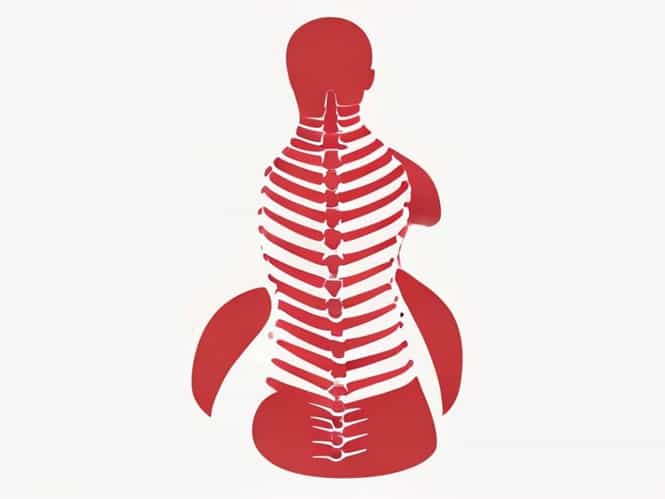
The erector spinae muscles are organized into three primary columns that run parallel along the spine from the sacrum and iliac crest to the skull. These muscles are divided based on their location and function, providing a framework for understanding their innervation.
Iliocostalis
- Located laterally within the erector spinae group.
- Extends from the iliac crest and sacrum to the ribs and cervical vertebrae.
- Assists in lateral flexion and extension of the spine.
Longissimus
- Intermediate column of the erector spinae.
- Extends from the lumbar region to the cervical vertebrae and mastoid process of the skull.
- Functions to extend the spine and head, as well as assist in lateral flexion.
Spinalis
- Medial column closest to the vertebral column.
- Runs along the spinous processes from the upper lumbar to the cervical region.
- Mainly responsible for extending the vertebral column and maintaining upright posture.
Innervation of the Erector Spinae Muscles
The erector spinae muscles are innervated primarily by the dorsal rami of the spinal nerves. These nerves provide motor signals that allow contraction and relaxation, as well as sensory input that communicates tension and position to the central nervous system. Understanding the segmental pattern of innervation is critical for both clinical assessments and interventions.
Dorsal Rami of Spinal Nerves
The dorsal rami are branches of the spinal nerves that emerge from the spinal cord and loop around to the posterior aspect of the body. They specifically innervate the deep muscles of the back, including the erector spinae, and provide sensation to the overlying skin. Each segment of the erector spinae is typically supplied by the dorsal ramus corresponding to that spinal level.
Segmental Distribution
- Cervical Region The dorsal rami of the cervical spinal nerves (C1-C8) innervate the upper portions of the erector spinae, including the cervical longissimus and spinalis muscles.
- Thoracic Region The thoracic dorsal rami (T1-T12) supply the thoracic portions of iliocostalis, longissimus, and spinalis, supporting extension and lateral movements of the thoracic spine.
- Lumbar Region The lumbar dorsal rami (L1-L5) provide innervation to the lower segments, including the lumbar iliocostalis and longissimus muscles, critical for lifting and stabilizing the lower back.
- Sacral Region The sacral dorsal rami contribute to the innervation of the sacral portions of the erector spinae, particularly affecting posture and pelvic stabilization.
Motor and Sensory Function
The dorsal rami carry both motor and sensory fibers. Motor fibers stimulate muscle contraction, enabling movement such as extension, lateral flexion, and rotation of the spine. Sensory fibers relay information about muscle stretch and tension, which helps the nervous system maintain balance and posture. Any disruption in this innervation can result in weakness, reduced mobility, or pain along the back.
Clinical Significance of Erector Spinae Innervation
Understanding the innervation of the erector spinae muscles is vital in clinical settings, particularly when assessing back pain, performing nerve blocks, or planning surgical procedures. Damage to the dorsal rami or their roots can manifest as motor deficits, sensory disturbances, or chronic pain.
Back Pain and Muscle Dysfunction
- Compression or injury to dorsal rami can lead to localized or radiating pain along the back.
- Weakness in the erector spinae muscles due to nerve impairment may result in poor posture, difficulty standing upright, or limited spinal mobility.
- Trigger points and myofascial pain syndromes in the erector spinae are often related to disrupted nerve signaling.
Surgical Considerations
During spinal surgeries, awareness of the dorsal rami pathways helps prevent accidental nerve damage. Surgeons must navigate around these nerves to preserve muscle function and minimize post-operative complications.
Rehabilitation and Physical Therapy
- Targeted exercises for the erector spinae aim to strengthen the muscles while ensuring proper nerve function.
- Electromyography (EMG) studies can assess innervation and muscle activation during therapy.
- Proper rehabilitation strategies reduce the risk of re-injury and improve recovery from nerve-related back conditions.
Testing and Assessing Erector Spinae Innervation
Healthcare providers can assess the function of the dorsal rami and the erector spinae muscles through various tests. Clinical evaluation often includes muscle strength testing, reflex assessment, and imaging studies to determine nerve integrity.
Electromyography (EMG)
EMG measures electrical activity in the muscles and can indicate whether the dorsal rami are effectively stimulating the erector spinae muscles. Abnormal results may suggest nerve compression, injury, or neuropathy.
Nerve Conduction Studies
- These studies evaluate the speed and quality of electrical signals traveling along the dorsal rami.
- Reduced conduction velocity may indicate nerve damage affecting the erector spinae muscles.
Physical Examination
Physicians assess posture, spinal alignment, and muscle tone to determine functional capacity. Weakness or asymmetry in the erector spinae may point to impaired innervation or nerve root compression.
The innervation of the erector spinae muscles is primarily provided by the dorsal rami of the spinal nerves, encompassing cervical, thoracic, lumbar, and sacral segments. This innervation allows precise control over spinal movements, maintaining posture, and enabling extension, lateral flexion, and rotation of the back. Understanding the anatomy and nerve supply of the erector spinae is essential for diagnosing back pain, planning surgical interventions, and developing effective rehabilitation programs. Clinical assessments such as EMG, nerve conduction studies, and physical examinations help evaluate the integrity of dorsal rami function. By recognizing the importance of proper innervation, healthcare professionals can ensure better patient outcomes, minimize the risk of chronic pain, and enhance the functional recovery of the spine and its associated musculature.