Breathing is a vital process that allows oxygen to enter the body and carbon dioxide to exit. This exchange of gases is made possible by a series of interconnected structures that form the upper and lower respiratory tract. The nasal cavity, pharynx, larynx, and trachea each play a distinct role in ensuring that air travels efficiently to the lungs. These anatomical parts not only transport air but also filter, warm, moisten, and protect the respiratory system. Understanding how the nasal cavity, pharynx, larynx, and trachea function together helps us appreciate the complexity of our respiratory health.
Nasal Cavity
Structure and Location
The nasal cavity is the large, air-filled space behind the nose. It is divided into two chambers by the nasal septum and is lined with a mucous membrane rich in blood vessels. The walls of the nasal cavity contain structures called nasal conchae or turbinates that increase the surface area for air to be processed.
Functions of the Nasal Cavity
- Air filtration: The nasal hairs and mucous trap dust, bacteria, and other foreign ptopics.
- Air humidification: Moisture from the mucous lining helps humidify the air before it reaches the lungs.
- Air warming: Blood vessels in the nasal cavity warm incoming air to body temperature.
- Smell: Olfactory receptors located in the upper part of the nasal cavity detect airborne chemicals, allowing the sense of smell.
Importance in Respiratory Health
By conditioning the air before it travels deeper into the respiratory system, the nasal cavity serves as the first line of defense. It protects the lungs from irritants, allergens, and pathogens, and plays a major role in respiratory immunity.
Pharynx
Structure and Subdivisions
The pharynx, commonly called the throat, is a muscular tube approximately 1214 cm long. It extends from the base of the skull to the level of the sixth cervical vertebra and is divided into three regions:
- Nasopharynx: Located behind the nasal cavity and above the soft palate.
- Oropharynx: Located behind the oral cavity and extends from the soft palate to the epiglottis.
- Laryngopharynx: The lower part of the pharynx that connects to the larynx and esophagus.
Functions of the Pharynx
The pharynx plays a dual role in both the respiratory and digestive systems:
- Passageway for air from the nasal cavity to the larynx
- Pathway for food and fluids to the esophagus
- Assists in equalizing air pressure in the middle ear through the Eustachian tubes
Muscle and Nerve Involvement
Muscles of the pharynx coordinate the swallowing process, preventing food from entering the respiratory tract. Innervation is mainly supplied by the glossopharyngeal and vagus nerves.
Larynx
Anatomy of the Larynx
The larynx, also known as the voice box, is located below the pharynx and above the trachea. It is composed of several cartilages, including the thyroid cartilage (Adam’s apple), cricoid cartilage, and epiglottis. The vocal cords are housed within the larynx.
Key Functions of the Larynx
- Voice production: The vocal cords vibrate to produce sound when air passes through them.
- Airway protection: During swallowing, the epiglottis folds down to cover the entrance to the trachea, preventing food or drink from entering the lungs.
- Breathing regulation: The muscles of the larynx adjust the size of the glottis (the space between vocal cords) to control airflow.
Role in Communication
The larynx enables human speech by working with the tongue, lips, and other parts of the vocal tract. Without the larynx, phonation sound production would not be possible.
Trachea
Structural Features
The trachea, or windpipe, is a tubular structure approximately 1012 cm long and about 2 cm in diameter. It extends from the larynx to the level of the fifth thoracic vertebra, where it splits into the left and right bronchi. The trachea is made up of C-shaped cartilaginous rings that provide structure and keep the airway open.
Functions of the Trachea
- Air conduction: The trachea provides a clear path for air to travel from the larynx to the bronchi and lungs.
- Mucociliary clearance: The tracheal lining contains cilia and mucus-producing cells that trap and remove ptopics and microbes.
- Structural support: The cartilage rings prevent collapse during breathing, especially when inhaling deeply.
Protection Mechanisms
If foreign objects enter the trachea, the body initiates a cough reflex. This powerful expulsion of air helps clear the airway and is a vital defense mechanism.
Relationship Between Nasal Cavity, Pharynx, Larynx, and Trachea
Sequential Airflow Pathway
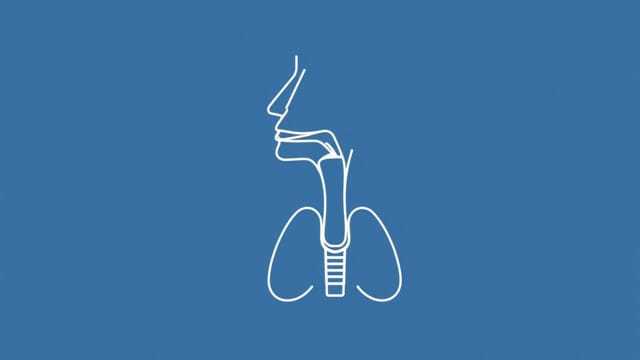
These four structures form a continuous airway:
- Air enters through the nasal cavity, where it is filtered, warmed, and humidified.
- It then passes into the pharynx, which acts as a shared pathway for both air and food.
- From there, air moves into the larynx, where voice production occurs and the airway is protected during swallowing.
- Finally, air travels down the trachea to reach the bronchi and eventually the lungs.
Coordinated Function
The nasal cavity, pharynx, larynx, and trachea work together in a highly coordinated manner. This coordination ensures that air flows freely, speech is possible, and that food and fluids do not accidentally enter the lungs. Any disruption in these structures due to injury, infection, or disease can significantly affect breathing and voice quality.
Common Conditions Affecting These Structures
Nasal Cavity Disorders
- Sinusitis
- Nasal polyps
- Deviated septum
Pharyngeal Conditions
- Pharyngitis (sore throat)
- Obstructive sleep apnea
- Tonsillitis
Laryngeal Issues
- Laryngitis
- Vocal cord nodules or polyps
- Laryngeal cancer
Tracheal Problems
- Tracheitis (inflammation)
- Tracheal stenosis (narrowing)
- Foreign body aspiration
The nasal cavity, pharynx, larynx, and trachea are essential components of the respiratory system. Each structure plays a unique and interdependent role in ensuring safe and efficient breathing, voice production, and protection of the lungs. Together, they form a well-coordinated airway that supports life and communication. Understanding their anatomy and function provides valuable insight into how our body manages one of its most critical tasks: respiration.