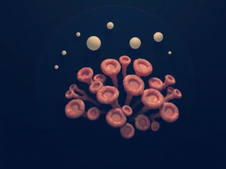
Porous coral-like outgrowths in the orbits are unusual clinical findings that can raise significant concern for both patients and healthcare professionals. These growths are characterized by a spongy, irregular, or branching appearance, resembling coral, and can affect the orbital bones surrounding the eyes. Such lesions may arise from benign or malignant conditions and can lead to symptoms such as proptosis, vision changes, or orbital discomfort. Understanding the causes, imaging characteristics, clinical manifestations, and management options is crucial for accurate diagnosis and effective treatment. Proper recognition of these outgrowths can also guide surgical planning and improve outcomes while minimizing complications.
Understanding Orbital Outgrowths
Orbital outgrowths can originate from bone, soft tissue, or a combination of both within the orbital cavity. The term porous coral-like typically refers to lesions with multiple small cavities or trabecular patterns that create a spongy radiographic or histological appearance. These outgrowths may be slow-growing or aggressive, depending on the underlying etiology. Their location in the orbit can lead to compression of the optic nerve, displacement of ocular structures, or cosmetic deformities, making timely diagnosis essential for preserving vision and orbital function.
Common Causes
Several conditions can result in porous, coral-like orbital outgrowths. They include
- Fibrous DysplasiaA benign bone disorder where normal bone is replaced with fibrous tissue, creating a characteristic ground glass or porous appearance on imaging. Orbital involvement can lead to proptosis and visual disturbances.
- OsteomasBenign bony tumors that may exhibit a coral-like structure. Osteomas of the orbital bones are rare but can cause cosmetic deformity or compression of surrounding structures.
- Osteogenic SarcomasMalignant bone tumors that sometimes present with irregular, spiculated, or porous growth patterns. Early detection is critical to prevent local invasion and metastasis.
- Paget’s Disease of BoneA disorder of bone remodeling that can lead to thickened, porous bones. Orbital involvement is uncommon but may result in craniofacial deformities.
- Multiple Myeloma or PlasmacytomaPlasma cell neoplasms can produce osteolytic lesions with a porous, punched-out appearance in the orbital bones.
- Metastatic LesionsCertain cancers, such as breast or prostate carcinoma, can metastasize to orbital bones, leading to lytic, spiculated, or porous changes visible on imaging.
Clinical Manifestations
The symptoms associated with porous coral-like outgrowths in the orbit vary depending on size, location, and rate of growth. Common clinical presentations include
- Proptosis or bulging of the eye, which may be unilateral or bilateral.
- Diplopia or double vision due to displacement of ocular muscles.
- Visual disturbances, including decreased acuity or field defects, if the optic nerve is compressed.
- Facial asymmetry or cosmetic changes, especially in slow-growing benign lesions.
- Pain or discomfort around the orbit, particularly in aggressive or inflammatory lesions.
- Occasionally, systemic symptoms such as fever or weight loss in the case of malignant or metastatic lesions.
Diagnostic Imaging
Imaging is essential for evaluating porous coral-like orbital outgrowths. Several modalities provide complementary information
- Computed Tomography (CT) ScanOffers excellent bone detail and can reveal porous, spiculated, or ground-glass appearances characteristic of certain lesions like fibrous dysplasia or osteomas. CT is particularly useful for surgical planning.
- Magnetic Resonance Imaging (MRI)Provides superior soft tissue contrast, helping to assess involvement of ocular muscles, optic nerve, and surrounding soft tissues. MRI is also valuable in differentiating benign from malignant lesions based on signal characteristics.
- X-rayCan demonstrate generalized bone changes but is less specific than CT or MRI. Plain radiographs may reveal a coarse, porous appearance in some bony lesions.
- Bone ScintigraphyUseful for identifying active bone lesions, particularly in fibrous dysplasia or Paget’s disease.
Histopathology
Definitive diagnosis often requires tissue sampling through biopsy. Histopathologic examination can reveal the cellular and structural characteristics of the lesion, including
- Fibrous tissue replacing normal bone in fibrous dysplasia.
- Mature lamellar bone in osteomas with branching or trabecular patterns.
- Atypical osteoid production or malignant cells in osteosarcomas.
- Plasma cell infiltration in multiple myeloma or plasmacytomas.
Management and Treatment
Treatment depends on the underlying cause, size, symptoms, and potential for vision impairment
- ObservationSmall, asymptomatic lesions may be monitored with periodic imaging, particularly benign lesions like slow-growing osteomas.
- Surgical InterventionIndicated for lesions causing proptosis, optic nerve compression, or cosmetic concerns. Complete excision may be required in malignant cases.
- Medical TherapyCertain conditions, such as Paget’s disease, may respond to bisphosphonates or other medications to stabilize bone remodeling.
- Radiation or ChemotherapyUsed for malignant lesions, metastatic disease, or plasma cell neoplasms affecting the orbital bones.
Prognosis
The prognosis of porous coral-like outgrowths in the orbits varies widely based on etiology. Benign lesions like fibrous dysplasia or osteomas generally have an excellent prognosis, with slow progression and minimal complications if monitored. Malignant lesions or aggressive bony tumors may have a guarded prognosis, particularly if diagnosis is delayed or if there is significant orbital or systemic involvement. Early detection, accurate imaging interpretation, and timely intervention are critical for preserving vision, preventing complications, and optimizing outcomes.
Key Points for Clinical Practice
- Always consider both benign and malignant causes when encountering coral-like orbital outgrowths.
- Use multimodal imaging, particularly CT and MRI, to assess the lesion’s extent, composition, and impact on orbital structures.
- Monitor asymptomatic lesions with periodic follow-up and imaging to detect progression.
- Engage a multidisciplinary team including ophthalmology, radiology, neurosurgery, and oncology for comprehensive management.
- Educate patients and families about potential symptoms, treatment options, and prognosis to ensure informed decision-making.
Porous coral-like outgrowths in the orbits represent a rare but clinically significant group of lesions that can affect vision, ocular function, and facial aesthetics. Recognizing their unique imaging characteristics, understanding potential etiologies, and applying appropriate diagnostic and management strategies are essential for optimal patient care. Through careful evaluation, timely intervention, and ongoing monitoring, healthcare providers can address the challenges posed by these lesions, protect vision, and improve overall quality of life for affected individuals. Multidisciplinary collaboration and patient-centered care are key elements in managing these complex orbital conditions effectively.