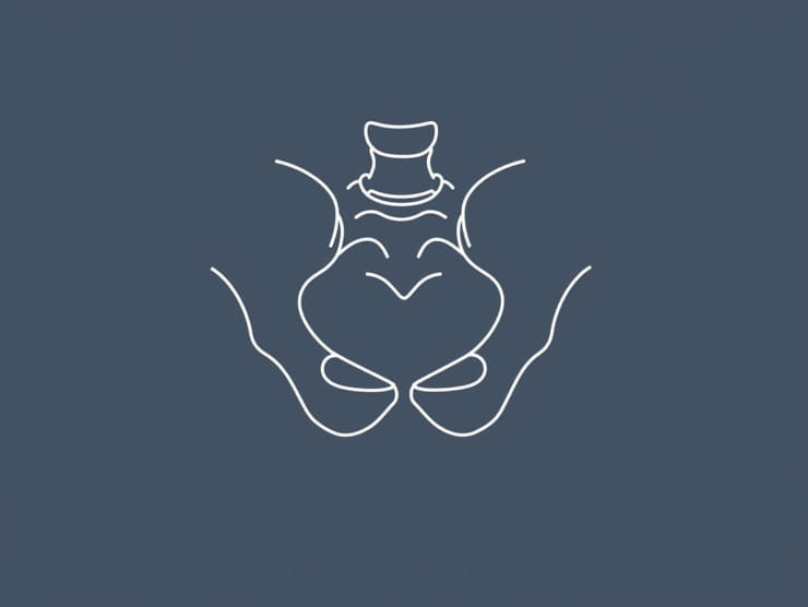
The tendon conjoint canal in the inguinal region is a critical anatomical structure that plays an essential role in the integrity of the lower abdominal wall. Understanding its location, composition, and clinical significance is vital for healthcare professionals, especially surgeons dealing with hernia repairs and other inguinal procedures. The tendon conjoint, also known as the conjoint tendon or inguinal aponeurotic falx, is formed by the fusion of the lower fibers of the internal oblique and transversus abdominis muscles. It contributes to the posterior wall of the inguinal canal and is a key landmark in preventing hernia recurrence during surgical interventions. Knowledge of its variations, attachments, and functional importance can aid in precise surgical planning and improve patient outcomes.
Anatomy of the Tendon Conjoint Canal
The tendon conjoint canal is located in the lower part of the anterior abdominal wall, just medial to the deep inguinal ring. Its main components include
- Internal oblique muscle fibersThese fibers run obliquely and contribute to the formation of the upper portion of the conjoint tendon.
- Transversus abdominis muscle fibersThe lower fibers of this muscle merge with the internal oblique to form the posterior reinforcement of the inguinal canal.
- Insertion pointThe conjoint tendon inserts primarily into the pubic crest and the pectineal line of the pubis, strengthening the medial aspect of the inguinal canal.
Function of the Tendon Conjoint Canal
The primary function of the tendon conjoint canal is to reinforce the posterior wall of the inguinal canal, particularly the medial region known as Hesselbach’s triangle. This area is a common site for direct inguinal hernias, and the tendon conjoint provides structural support to prevent abdominal contents from protruding. Additionally, it acts as a stabilizing structure during increased intra-abdominal pressure, such as coughing, sneezing, or straining.
Clinical Significance
Understanding the tendon conjoint canal is crucial for both diagnosis and surgical management of inguinal hernias. Key clinical points include
- Hernia repairDuring open or laparoscopic inguinal hernia repair, identification of the conjoint tendon is essential to reinforce the posterior wall properly and reduce recurrence.
- Direct herniasA weak or attenuated conjoint tendon can predispose individuals to direct inguinal hernias, which protrude through Hesselbach’s triangle.
- Surgical landmarksThe tendon conjoint serves as an important anatomical landmark during inguinal procedures, guiding safe dissection and mesh placement.
- Anatomic variationsThe size, thickness, and composition of the tendon conjoint can vary between individuals, influencing surgical approaches and outcomes.
Relationship with Surrounding Structures
The tendon conjoint canal is closely related to several important anatomical structures
- Inguinal ligamentThe conjoint tendon lies immediately superior and medial to the inguinal ligament, contributing to the lower abdominal wall’s structural integrity.
- Hesselbach’s triangleThis triangular area is bordered by the inferior epigastric vessels laterally, the lateral border of the rectus abdominis medially, and the inguinal ligament inferiorly. The tendon conjoint reinforces the posterior wall here.
- Deep inguinal ringLocated lateral to the conjoint tendon, the deep inguinal ring serves as the entrance for structures of the spermatic cord in males or the round ligament in females.
- Spermatic cord and round ligamentThese structures pass through the inguinal canal and are closely associated with the tendon conjoint, emphasizing the importance of careful dissection during surgery.
Variations in Anatomy
The anatomy of the tendon conjoint canal can exhibit variations that may impact surgical strategies
- ThicknessSome individuals have a robust conjoint tendon, while others may have a thin or attenuated structure, increasing hernia risk.
- LengthVariability in tendon length can affect the degree of reinforcement provided to the posterior wall.
- Muscle fiber orientationDifferences in fiber alignment may influence surgical technique during hernia repair.
Surgical Relevance
Inguinal hernia repair, particularly the Lichtenstein tension-free method and laparoscopic approaches, requires precise understanding of the tendon conjoint canal
- Mesh placementProper identification of the conjoint tendon ensures correct mesh positioning and secure fixation, reducing recurrence.
- Direct hernia repairReinforcement of the weakened posterior wall with the conjoint tendon is a key step in direct hernia repair.
- Preservation of structuresKnowledge of the tendon’s relationship with the spermatic cord, vessels, and nerves prevents inadvertent injury.
- Minimizing complicationsAwareness of anatomical variations allows surgeons to tailor techniques and avoid complications such as chronic pain or recurrence.
Imaging and Diagnosis
While the tendon conjoint canal is primarily of surgical interest, imaging modalities can help in assessing its integrity, particularly in recurrent hernias or atypical presentations
- Ultrasound Can visualize the inguinal canal and assess the integrity of the posterior wall.
- CT scan Provides detailed anatomy and helps in planning complex hernia repairs.
- MRI Useful in evaluating soft tissue structures and identifying anatomical variations.
Complications of Tendon Conjoint Weakness
Weakness or defects in the tendon conjoint canal can lead to clinical issues, primarily related to hernia formation
- Direct inguinal hernias Protrusion of abdominal contents through Hesselbach’s triangle due to insufficient reinforcement.
- Recurrent hernias Incomplete repair or unnoticed variations can predispose to recurrence after surgery.
- Postoperative complications Inadequate identification of the tendon may result in improper mesh placement or injury to adjacent structures.
The tendon conjoint canal in the inguinal region is a crucial anatomical structure that reinforces the lower abdominal wall and plays a key role in hernia prevention. Understanding its anatomy, variations, and relationship with surrounding structures is essential for clinicians, especially surgeons involved in inguinal hernia repair. Proper identification and reinforcement of the conjoint tendon during surgical procedures help minimize complications, prevent hernia recurrence, and improve patient outcomes. Continuous research and detailed anatomical studies further enhance our understanding of this important structure, ensuring safe and effective surgical interventions for patients with inguinal pathologies.