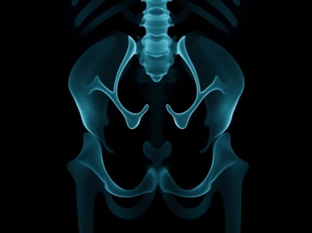
Urethral injuries can occur due to trauma, surgical complications, or medical conditions affecting the urinary tract. Diagnosing these injuries accurately is crucial for preventing long-term complications such as urinary incontinence, strictures, or infection. One of the primary diagnostic tools used in evaluating urethral injuries is X-ray imaging, often combined with contrast studies to provide detailed visualization of the urethra and surrounding structures. Understanding how urethral injury X-rays work, when they are indicated, and what findings can be expected is essential for both medical professionals and patients.
What is a Urethral Injury?
A urethral injury refers to damage to the urethra, the tube responsible for transporting urine from the bladder to the outside of the body. Injuries can range from minor contusions to complete disruptions, often classified based on location as anterior or posterior urethral injuries. Anterior injuries typically involve the penile or bulbar urethra, while posterior injuries affect the membranous or prostatic urethra. The mechanism of injury can include blunt trauma, pelvic fractures, straddle injuries, catheterization, or iatrogenic causes during medical procedures.
Symptoms of Urethral Injury
Patients with urethral injury may present with a variety of symptoms depending on the severity and location of the damage
- Painful urination or difficulty voiding
- Blood at the urethral opening (urethral bleeding)
- Inability to pass urine
- Swelling, bruising, or hematoma in the genital or perineal area
- Signs of pelvic trauma, particularly in posterior urethral injuries
The Role of X-Ray Imaging in Urethral Injuries
X-ray imaging plays a pivotal role in diagnosing urethral injuries, particularly when used in combination with contrast media. The most common radiologic procedure is the retrograde urethrogram (RUG), which involves the introduction of a contrast agent into the urethra to highlight any disruption or stricture on X-ray images. This approach allows clinicians to identify the exact location, extent, and type of urethral injury.
Retrograde Urethrogram (RUG)
The retrograde urethrogram is considered the gold standard for evaluating urethral injuries. The procedure involves the following steps
- The patient is positioned appropriately, often lying on their back with legs slightly apart.
- A catheter or syringe is used to gently introduce contrast dye into the distal urethra.
- X-ray images are taken while the contrast flows through the urethra, highlighting any leaks, strictures, or disruptions.
- Images are reviewed to assess the site and severity of the injury, guiding further management and surgical planning.
Advantages of X-Ray Evaluation
X-ray imaging, particularly with contrast, offers several advantages for assessing urethral injuries
- Non-invasive visualization of urethral anatomy and injury sites
- Accurate determination of injury location and length
- Identification of associated complications such as extravasation of urine or contrast into surrounding tissues
- Facilitation of treatment planning, including surgical repair or catheter placement
Other Imaging Considerations
In some cases, additional imaging may complement X-ray studies
- Voiding Cystourethrogram (VCUG)Used to assess the urethra during urination, particularly useful for detecting posterior injuries or bladder involvement.
- CT or MRI ScansMay be employed for complex trauma cases to evaluate surrounding pelvic structures or identify concurrent injuries.
Interpreting Urethral X-Ray Findings
Radiologists and urologists look for several key indicators when reviewing urethral injury X-rays
- Partial TearContrast leaks partially outside the urethral lumen, indicating an incomplete disruption.
- Complete DisruptionContrast fails to pass beyond the injury site, suggesting a full transection of the urethra.
- StricturesNarrowing of the urethral lumen caused by scar tissue or chronic injury.
- ExtravasationLeakage of urine or contrast into surrounding tissues, often seen as a darkened area outside the urethral path.
Management Based on X-Ray Findings
The treatment of urethral injuries depends on the severity and location identified by X-ray studies. Management strategies may include
- CatheterizationPlacement of a urethral or suprapubic catheter to allow urinary drainage while the injury heals.
- Surgical RepairUrethroplasty or other surgical interventions for complete disruptions or complex injuries.
- ObservationMinor partial tears may heal with conservative management, including catheterization and close monitoring.
- Follow-Up ImagingRepeat X-rays or contrast studies may be performed to assess healing progress or detect complications.
Complications and Risks
Although X-ray imaging with contrast is generally safe, potential risks include
- Allergic reactions to contrast material
- Discomfort during catheter insertion or contrast injection
- Radiation exposure, which is minimal but considered in repeated studies
- Infection, particularly if sterility is not maintained during the procedure
Urethral injury X-rays, particularly retrograde urethrograms, provide a vital diagnostic tool for accurately assessing trauma or damage to the urethra. By offering detailed visualization of the urethral lumen, injury location, and any associated complications, these imaging studies allow healthcare professionals to plan effective treatment strategies. Timely and accurate diagnosis is critical in preventing long-term complications such as strictures, incontinence, or recurrent infections. Understanding the role, procedure, and interpretation of urethral injury X-rays helps both medical practitioners and patients navigate this complex area of urologic care with confidence, ensuring optimal outcomes and supporting overall urinary tract health.