Groin injuries are common, especially among athletes, physically active individuals, and those involved in sudden movements or trauma. These injuries can range from minor muscle strains to more serious conditions like fractures, hernias, or hip joint pathology. X-ray imaging plays an essential role in the diagnosis and management of groin injuries, providing clear visualization of bones, joints, and certain soft tissue changes. By understanding when and how X-rays are used, patients and healthcare providers can ensure timely and accurate assessment, leading to appropriate treatment and faster recovery.
Understanding Groin Injuries
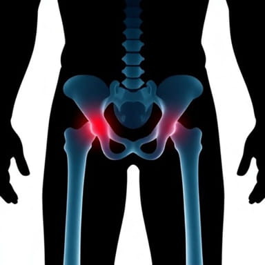
The groin region is a complex area composed of muscles, tendons, ligaments, bones, and joints. Common injuries include muscle strains, ligament sprains, hip joint injuries, pubic bone fractures, and hernias. Symptoms often include pain, swelling, bruising, limited mobility, and difficulty bearing weight. While physical examination is crucial, imaging like X-rays provides additional information about underlying structural damage that might not be evident through clinical assessment alone.
When X-Ray Imaging is Recommended
X-rays are generally recommended for groin injuries when there is suspicion of bone involvement, persistent pain despite initial treatment, or trauma resulting in severe discomfort. Specific scenarios include
- Suspected fractures of the pelvis, pubic bone, or femur.
- Evaluation of hip joint dislocations or subluxations.
- Assessment of bone abnormalities or degenerative changes in chronic groin pain.
- Pre-surgical planning for orthopedic interventions.
- Monitoring healing progress after previous fractures or surgery.
X-Ray Technique for Groin Injury
Performing an X-ray for a groin injury requires proper positioning and technique to ensure accurate imaging. Standard views may include anteroposterior (AP), lateral, and oblique projections. Each projection helps visualize different aspects of the hip joint, pelvic bones, and femur.
Anteroposterior (AP) View
In the AP view, the patient lies on their back with legs extended. The X-ray beam passes from front to back, providing a clear image of the pelvis, hip joints, and proximal femur. This view is essential for detecting fractures, dislocations, and bone alignment abnormalities.
Lateral View
The lateral view involves positioning the patient on their side with the affected hip facing upward. This projection provides a side profile of the femur, hip joint, and pelvic bones, helping detect subtle fractures or displacement not visible on the AP view.
Oblique Views
Oblique views may be taken by slightly rotating the patient’s pelvis or leg to highlight specific areas, such as the acetabulum, pubic rami, or iliac bones. These views are particularly useful for identifying complex fractures or evaluating bone alignment in multiple planes.
Patient Preparation and Safety
Before undergoing an X-ray for groin injuries, patients should remove clothing or metallic objects near the pelvic area, such as belts, buttons, or zippers. Female patients may be asked to wear a lead apron to protect reproductive organs from unnecessary radiation. The procedure is quick, non-invasive, and generally safe, with minimal radiation exposure compared to CT scans.
Radiographic Findings
X-ray imaging provides valuable information regarding the structural integrity of bones and joints. Key findings include
Fractures
Fractures in the pelvis, pubic rami, or femoral neck can be visualized as breaks or lines in the bone. The location, pattern, and severity of fractures determine treatment options, which may include rest, immobilization, or surgical intervention.
Dislocations
Hip or pelvic joint dislocations can be identified by abnormal alignment of bones. X-rays help assess the degree of displacement and guide orthopedic management.
Bone Degeneration and Osteoarthritis
Chronic groin pain may be due to degenerative changes in the hip joint or surrounding bones. X-rays reveal narrowing of joint spaces, osteophyte formation, and other signs of osteoarthritis, which assist in long-term management planning.
Foreign Bodies or Abnormalities
In cases of trauma, X-rays can detect foreign objects or unusual bone growths that may contribute to groin pain or complicate treatment.
Advantages of X-Ray in Groin Injury Assessment
- Quick and widely available diagnostic tool.
- Effective in detecting fractures, dislocations, and bone abnormalities.
- Low radiation exposure compared to advanced imaging like CT scans.
- Supports treatment planning and monitoring of healing progress.
Limitations of X-Ray Imaging
While X-rays are excellent for visualizing bone structures, they have limited ability to detect soft tissue injuries such as muscle strains, ligament tears, or tendon injuries. In such cases, additional imaging like MRI or ultrasound may be necessary. X-rays may also miss subtle hairline fractures or stress fractures, particularly in the early stages, making clinical correlation essential.
Complementary Imaging Modalities
Magnetic Resonance Imaging (MRI)
MRI provides detailed images of soft tissues, including muscles, tendons, ligaments, and cartilage. It is often used when X-rays are inconclusive or when soft tissue injury is suspected alongside bone involvement.
Computed Tomography (CT)
CT scans offer high-resolution cross-sectional images of bones and joints, ideal for complex fractures, dislocations, or pre-surgical evaluation. CT can also detect small fragments that might be missed on X-rays.
Ultrasound
Ultrasound is useful for evaluating soft tissue injuries, hematomas, and hernias in the groin region. It is safe, non-invasive, and can be performed bedside, making it valuable in emergency situations.
Management Based on X-Ray Findings
Treatment strategies depend on the specific injury identified through X-ray imaging
- FracturesMay require rest, immobilization, or surgical fixation depending on severity.
- DislocationsOften require urgent reduction by a trained orthopedic specialist.
- Bone abnormalities or osteoarthritisManaged with physical therapy, medication, or surgical intervention if conservative measures fail.
- Soft tissue injury suspicionFurther imaging with MRI or ultrasound may guide rehabilitation or surgical repair.
X-ray imaging is a fundamental tool in the evaluation of groin injuries, providing essential information about bone integrity, joint alignment, and potential fractures or dislocations. Proper patient positioning, technique, and interpretation are crucial for accurate diagnosis. While X-rays have limitations in visualizing soft tissues, they remain a quick, cost-effective, and widely accessible method for initial assessment. Complementary imaging modalities, such as MRI, CT, or ultrasound, may be required in complex cases. By combining clinical evaluation with radiographic findings, healthcare providers can implement appropriate treatment plans, ensure optimal recovery, and minimize long-term complications for patients with groin injuries.