The human torso is a complex anatomical region that houses critical organs, bones, muscles, and blood vessels. When patients experience trauma, pain, or unexplained symptoms in this area, X-ray imaging is one of the first diagnostic tools utilized to assess the internal structure. X-rays of the torso provide a detailed view of the chest, abdomen, spine, and surrounding tissues, allowing healthcare providers to identify fractures, organ abnormalities, and other pathologies. Understanding the purpose, technique, and interpretation of torso X-rays is essential for accurate diagnosis and timely treatment, helping to prevent complications and improve patient outcomes.
Understanding the Human Torso
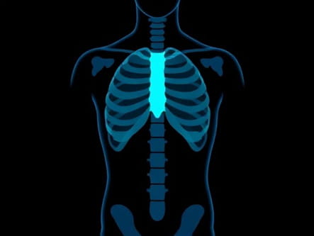
The human torso extends from the neck down to the pelvis and includes the thoracic cage, spine, abdominal cavity, and pelvis. It contains vital organs such as the heart, lungs, liver, stomach, intestines, and kidneys. Additionally, the rib cage protects the thoracic organs while supporting respiration, and the spine provides structural support and houses the spinal cord. Given this complexity, injuries or diseases in the torso can have significant health implications, making accurate imaging crucial.
Common Indications for Torso X-Ray
X-rays of the torso are typically requested for various clinical situations. Some common indications include
- Suspected fractures of the ribs, sternum, or vertebrae after trauma or falls.
- Evaluation of chest pain, shortness of breath, or suspected lung disease.
- Assessment of abdominal pain, bowel obstruction, or organ enlargement.
- Pre-surgical planning for spinal, thoracic, or abdominal procedures.
- Monitoring chronic conditions such as scoliosis or degenerative spinal disease.
Techniques for Torso X-Ray Imaging
Proper technique is essential to obtain accurate and high-quality X-ray images of the torso. Radiographers use multiple views to visualize different structures and detect abnormalities.
Chest X-Ray
The chest X-ray is one of the most common imaging studies for the torso. It typically involves an anteroposterior (AP) or posteroanterior (PA) view, along with a lateral view. Patients are asked to stand or sit upright, take a deep breath, and hold it briefly to expand the lungs and improve image clarity. Chest X-rays reveal the lungs, heart, major blood vessels, and bones of the thoracic cage.
Abdominal X-Ray
Abdominal X-rays are used to evaluate the organs, bowel, and surrounding structures. Images are usually taken in an erect or supine position to detect air-fluid levels, organ enlargement, or foreign bodies. In some cases, multiple views may be needed to provide a complete assessment.
Spinal X-Ray
Spinal X-rays assess the vertebrae in the cervical, thoracic, and lumbar regions. Proper positioning is critical to detect fractures, misalignments, or degenerative changes. Standard views include AP, lateral, and oblique projections.
Preparation and Safety
Before undergoing a torso X-ray, patients may need to remove clothing, jewelry, or metallic objects that could interfere with imaging. Lead aprons are often used to protect reproductive organs and minimize radiation exposure. While X-rays involve low levels of radiation, proper safety measures are essential to reduce unnecessary exposure, especially in children and pregnant women.
Radiographic Findings in Torso X-Ray
X-rays of the torso can reveal a wide range of abnormalities. Understanding the typical findings is essential for accurate diagnosis and treatment planning.
Bone Fractures and Dislocations
Fractures of the ribs, clavicle, sternum, or vertebrae appear as lines or breaks in the bone. Dislocations, particularly in the spine or shoulder region, are identified by misalignment of bones. These findings are critical in trauma assessment and guiding orthopedic interventions.
Lung and Heart Abnormalities
Chest X-rays can detect pneumonia, lung masses, pleural effusions, or pneumothorax. Heart enlargement, congestive heart failure, or major vessel abnormalities are also identifiable. Accurate interpretation helps in diagnosing acute or chronic cardiopulmonary conditions.
Abdominal Organ Assessment
Abdominal X-rays can reveal bowel obstruction, kidney stones, or abnormal gas patterns. Organ enlargement, calcifications, or foreign bodies are also detectable. These findings aid in planning further imaging, treatment, or surgical intervention.
Spinal and Skeletal Changes
Spinal X-rays provide insight into scoliosis, kyphosis, degenerative disc disease, or vertebral fractures. Subtle misalignments or bone density changes can be critical in managing chronic pain or planning surgery.
Advantages of Torso X-Ray
- Non-invasive and widely available diagnostic tool.
- Quick imaging procedure with immediate results.
- Effective for detecting fractures, organ enlargement, and chest pathology.
- Supports clinical decision-making and treatment planning.
Limitations of Torso X-Ray
Despite its advantages, X-ray imaging has limitations. It has low sensitivity for soft tissue injuries, subtle organ pathology, or early-stage disease. For detailed assessment of organs, muscles, blood vessels, or tumors, additional imaging modalities such as CT scans, MRI, or ultrasound may be necessary. Moreover, overlapping structures in the torso can sometimes obscure abnormalities.
Complementary Imaging Modalities
Computed Tomography (CT)
CT scans provide detailed cross-sectional images of the torso, allowing better visualization of bones, organs, and soft tissues. They are particularly useful in trauma, tumor assessment, and complex fractures.
Magnetic Resonance Imaging (MRI)
MRI offers high-resolution images of soft tissues, spinal cord, muscles, and organs without radiation exposure. It is used when X-rays are inconclusive or when soft tissue evaluation is needed.
Ultrasound
Ultrasound is ideal for evaluating abdominal organs, detecting fluid collections, or guiding interventional procedures. It is safe, portable, and non-invasive.
Clinical Importance
X-rays of the human torso are crucial for the initial assessment of injuries, chronic conditions, or unexplained symptoms. They help clinicians make informed decisions about treatment, identify urgent issues requiring intervention, and monitor recovery or progression of disease. Combining X-ray findings with physical examination and patient history ensures accurate diagnosis and optimal patient care.
X-ray imaging of the human torso is a fundamental diagnostic tool that provides vital information about bones, joints, and certain internal organs. With proper technique, positioning, and interpretation, X-rays can detect fractures, dislocations, organ abnormalities, and chronic conditions, guiding appropriate medical or surgical management. While limited in evaluating soft tissue pathology, X-rays remain a quick, accessible, and cost-effective method for initial assessment. When combined with complementary imaging such as CT, MRI, or ultrasound, torso X-rays contribute significantly to comprehensive patient care, ensuring timely diagnosis, effective treatment, and improved outcomes for individuals experiencing torso-related injuries or medical conditions.