An X-ray of the ureter is a specialized medical imaging procedure that helps doctors visualize the urinary tract and diagnose conditions affecting the ureters. The ureters are tubes that carry urine from the kidneys to the bladder, and issues in these structures can cause pain, obstruction, or infection. Understanding the correct term for an X-ray of the ureter, how it is performed, and its medical significance is essential for students, healthcare professionals, and patients seeking information about diagnostic procedures. This topic explores the procedure, its indications, preparation, and interpretation, providing a comprehensive overview of ureter imaging in modern medicine.
Definition and Terminology
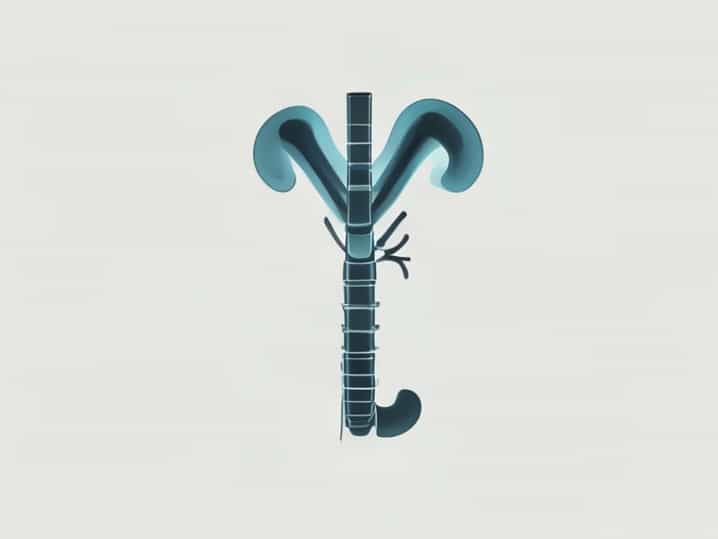
An X-ray of the ureter is called anintravenous pyelogram (IVP)or aurogram. This imaging technique uses contrast material injected into the bloodstream to make the urinary tract visible on X-ray films. The contrast highlights the kidneys, ureters, and bladder, allowing physicians to identify blockages, stones, tumors, or other abnormalities. While modern imaging techniques like CT urography or MRI are becoming more common, the IVP remains a valuable tool in many healthcare settings due to its effectiveness and relatively low cost.
Purpose of Ureter Imaging
Imaging the ureters with an X-ray serves several important purposes
- Detecting kidney stones that may be obstructing the ureters.
- Evaluating congenital abnormalities in the urinary tract.
- Identifying strictures, narrowing, or blockages in the ureters.
- Assessing tumors or masses within the ureters or adjacent tissues.
- Monitoring post-surgical healing or complications in the urinary tract.
How an Intravenous Pyelogram is Performed
The procedure for obtaining an X-ray of the ureter involves several steps to ensure clear imaging and patient safety. IVP is typically performed in a radiology department under professional supervision. The steps include
1. Patient Preparation
Patients may be instructed to fast for several hours before the procedure and may receive a mild laxative to clear the intestines. This ensures that the urinary tract is not obscured by bowel contents. Patients should inform the doctor of any allergies, especially to iodine-based contrast material, kidney problems, or other medical conditions.
2. Administration of Contrast
A contrast dye, usually iodine-based, is injected into a vein, often in the arm. The dye travels through the bloodstream to the kidneys, where it is filtered into the urine. This makes the ureters and other parts of the urinary system visible on X-ray films.
3. Taking X-ray Images
After contrast injection, a series of X-rays is taken at different time intervals. Early images capture the dye entering the kidneys, while later images show it moving through the ureters and into the bladder. The radiologist monitors the flow of the contrast to detect abnormalities in shape, size, or function of the ureters.
4. Post-Procedure Care
After the procedure, patients are encouraged to drink plenty of fluids to help flush the contrast material from the body. Some mild side effects such as nausea or a warm sensation may occur during the injection. Severe reactions are rare but require immediate medical attention.
Indications for an X-ray of the Ureter
Doctors may recommend a ureter X-ray for several medical indications. Common reasons include
- Persistent or severe flank pain suggestive of kidney stones.
- Hematuria, or blood in the urine, which may indicate urinary tract injury, stones, or tumors.
- Recurrent urinary tract infections that could be caused by structural abnormalities.
- Evaluation of urinary tract function after surgery or trauma.
- Monitoring known conditions such as strictures or congenital ureter abnormalities.
Advantages of Ureter Imaging
An X-ray of the ureter provides several benefits over other diagnostic methods. It is relatively quick, widely available, and cost-effective compared to advanced imaging techniques. IVP can provide both structural and functional information, showing how urine flows through the kidneys and ureters. This combination helps doctors make accurate diagnoses and plan appropriate treatments for patients with urinary tract disorders.
Limitations
While useful, IVP has some limitations. It exposes the patient to a small amount of radiation and may not detect very small stones or early-stage tumors. Additionally, patients with severe kidney impairment may not be able to safely receive iodine-based contrast. In such cases, alternative imaging techniques like ultrasound, CT urography, or MRI may be recommended.
Interpreting the X-ray Results
Radiologists analyze the X-ray images to evaluate the ureters for size, shape, and patency. Key observations include
- Obstructions, which may appear as narrowed or blocked sections of the ureter.
- Stones, seen as dense white areas within the ureters.
- Structural abnormalities, such as strictures, duplications, or congenital malformations.
- Signs of infection or inflammation, indicated by changes in the lining or surrounding tissues.
- Abnormal masses or tumors, which may distort normal ureter anatomy.
Radiologists provide a detailed report to the referring physician, who then determines the appropriate treatment plan.
Modern Alternatives
While IVP has been a traditional method for imaging the ureters, modern alternatives offer enhanced imaging with fewer limitations. These include
- CT UrographyProvides highly detailed images of the urinary tract and can detect smaller stones and tumors.
- MRI UrographyUseful for patients who cannot receive iodine-based contrast and offers excellent soft tissue contrast.
- UltrasoundNon-invasive and radiation-free, useful for detecting stones and hydronephrosis but less effective for small ureteral abnormalities.
An X-ray of the ureter, known as an intravenous pyelogram or urography, is an important diagnostic tool in medicine for evaluating the urinary tract. It helps detect stones, blockages, tumors, and structural abnormalities, guiding treatment and management decisions. The procedure involves contrast injection, timed X-ray imaging, and careful analysis by radiologists. While modern techniques like CT and MRI urography are gaining popularity, IVP remains a valuable and widely used method in many clinical settings. Understanding this procedure, its indications, advantages, and limitations is essential for healthcare professionals and patients seeking clarity on urinary tract diagnostics.