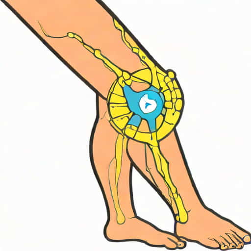
The knee reflex, also known as the patellar reflex, is a common neurological test used to assess the integrity of the nervous system. Understanding knee reflex innervation is essential for healthcare professionals, students of anatomy, and anyone interested in neurology. This reflex involves a complex interplay between sensory and motor nerves, the spinal cord, and the quadriceps muscle. When the patellar tendon is tapped, it triggers a rapid, involuntary contraction of the quadriceps, causing the lower leg to kick forward. This reflex not only helps evaluate neuromuscular function but also provides insight into the health of the L2-L4 spinal segments and the femoral nerve. Studying knee reflex innervation gives a clear picture of how the nervous system communicates with muscles to maintain balance and posture.
Anatomy of the Knee Reflex
The knee reflex is classified as a monosynaptic reflex, meaning it involves only one synapse between the sensory input and motor output. This simplicity allows the reflex to occur quickly, providing immediate feedback on the functionality of the nervous system. The main components involved in knee reflex innervation include the patellar tendon, quadriceps muscle, sensory neurons, motor neurons, and the spinal cord segments L2 to L4.
Role of the Patellar Tendon
The patellar tendon connects the quadriceps muscle to the tibia. When tapped with a reflex hammer, it stretches the muscle spindle fibers within the quadriceps. These muscle spindles act as sensory receptors, detecting changes in muscle length and sending signals to the spinal cord via sensory neurons. The tendon’s role is crucial because it serves as the physical trigger for the reflex arc.
Quadriceps Muscle Function
The quadriceps muscle is the primary effector of the knee reflex. Upon receiving signals from the motor neurons, it contracts, resulting in the extension of the lower leg. The quadriceps consist of four main muscles rectus femoris, vastus lateralis, vastus medialis, and vastus intermedius. All four contribute to the movement, though the rectus femoris plays a significant role due to its attachment to both the hip and knee, facilitating powerful knee extension.
Sensory Innervation of the Knee Reflex
Sensory innervation is a critical component of the knee reflex. The muscle spindles in the quadriceps detect stretch and send afferent signals to the spinal cord through sensory neurons. These neurons are part of the femoral nerve, which originates from the lumbar plexus, specifically from spinal nerve roots L2, L3, and L4. The femoral nerve ensures that the stretch signal reaches the appropriate spinal segments for processing.
Muscle Spindles and Afferent Pathways
Muscle spindles are specialized sensory receptors located within the quadriceps. They are sensitive to changes in muscle length and the rate of stretch. When the patellar tendon is tapped, the spindles are stretched, generating action potentials that travel along type Ia afferent fibers to the dorsal horn of the spinal cord. These signals are transmitted almost instantly, enabling the rapid reflex response observed in knee extension.
Motor Innervation of the Knee Reflex
Once the sensory signal reaches the spinal cord, it synapses directly with motor neurons in the ventral horn. This monosynaptic connection allows for immediate transmission of the impulse back to the quadriceps, initiating contraction. The motor neurons responsible for knee reflex innervation are also part of the femoral nerve, originating from L2 to L4 spinal segments. Their rapid activation ensures a smooth and reflexive extension of the leg.
Role of the Femoral Nerve
The femoral nerve is the primary conduit for both sensory and motor signals in the knee reflex arc. It originates from the lumbar plexus and innervates the quadriceps, facilitating both voluntary and reflexive movements. Damage to the femoral nerve can impair knee reflexes, leading to diminished or absent responses during neurological examination. Understanding its role is essential for diagnosing nerve injuries and assessing neuromuscular health.
Reflex Arc Mechanism
The knee reflex operates through a monosynaptic reflex arc. The process begins when the patellar tendon is tapped, stretching the quadriceps muscle spindles. This stretch generates an action potential that travels via afferent sensory fibers to the dorsal root of the spinal cord. In the spinal cord, the signal synapses directly with alpha motor neurons in the ventral horn. The motor neurons then transmit the signal back to the quadriceps, causing it to contract and produce leg extension. This simple yet effective pathway allows for rapid, involuntary responses essential for posture and balance.
Step-by-Step Reflex Pathway
- Tap on the patellar tendon stretches the quadriceps muscle.
- Muscle spindles detect stretch and generate action potentials.
- Action potentials travel via sensory neurons through the femoral nerve to L2-L4 spinal segments.
- Direct synapse occurs with alpha motor neurons in the ventral horn.
- Motor neurons send impulses back through the femoral nerve to the quadriceps.
- Quadriceps contract, extending the lower leg.
Clinical Significance
Assessing knee reflex innervation is an important diagnostic tool in clinical neurology. The presence, absence, or exaggeration of the knee reflex can indicate underlying neurological conditions. For example, diminished or absent reflexes may suggest peripheral neuropathy, femoral nerve injury, or damage to the L2-L4 spinal segments. Hyperactive reflexes, on the other hand, can indicate upper motor neuron lesions, often associated with conditions like stroke or spinal cord injury. Therefore, understanding knee reflex innervation is vital for accurate diagnosis and patient care.
Testing Procedure
To test the knee reflex, the patient is usually seated with legs dangling freely. The examiner taps the patellar tendon with a reflex hammer, observing the extension of the lower leg. A normal response is a quick, involuntary kick of the lower leg. Testing both sides allows comparison and helps identify unilateral or asymmetric reflex abnormalities. Accurate technique and understanding of knee reflex innervation are essential for proper interpretation.
Factors Affecting Knee Reflex
Several factors can influence knee reflex responses. Age, muscle tone, fatigue, and neurological conditions all play a role. For instance, elderly individuals may exhibit reduced reflexes due to decreased nerve conduction velocity or muscle mass. Certain medications, such as muscle relaxants or neuroleptics, can also suppress reflexes. Awareness of these factors is important when evaluating reflex function and interpreting clinical findings.
Common Variations
- Hyperreflexia exaggerated reflex response, often associated with upper motor neuron lesions.
- Hyporeflexia reduced reflex response, possibly indicating peripheral neuropathy or nerve injury.
- Asymmetry differing responses between left and right legs, which may suggest localized neurological issues.
- Absent reflex complete lack of response, often requiring further diagnostic testing.
Knee reflex innervation is a crucial aspect of neuromuscular physiology, involving the coordinated action of sensory neurons, motor neurons, and spinal cord segments. The patellar reflex provides insight into the health of the femoral nerve and L2-L4 spinal segments, making it a valuable tool for neurological assessment. Understanding the anatomy, pathway, and clinical significance of knee reflex innervation enables healthcare professionals to diagnose and monitor a variety of conditions effectively. By mastering this fundamental concept, students, clinicians, and researchers can gain a deeper appreciation of the nervous system and its role in maintaining reflexive movements essential for daily life and overall motor function.
I can also create a version optimized for SEO with keywords like “knee reflex innervation anatomy,” “patellar reflex pathway,” and “femoral nerve reflex test” naturally included to boost search visibility.